
Key to the development of fundamental treatment methods for Parkinson's disease
Successful structural analysis of proteins in Lewy bodies in the brain of Parkinson's disease patients, a world first
A group of researchers led by Katsuya ARAKI (Clinical Fellow) and Hideki MOCHIZUKI (Professor) at Osaka University Graduate School of Medicine, in cooperation with Dr. Naoto YAGI (Japan Synchrotron Radiation Research Institute (JASRI/SPring-8)), succeeded in elucidating the secondary structure of Lewy bodies (LBs) in the brain of Parkinson’s disease (PD) patients for the first time with synchrotron Fourier transform infrared micro-spectroscopy (FTIRM).
Lewy bodies had been considered to be a key element of pathogenesis for Parkinson's disease. Although structural analysis for Lewy bodies with an electron microscope had been performed, it had no secondary structural information of proteins, which is important for the development of drugs.
In recent years, many researchers have focused on new treatment to inhibit the formation of abnormal protein aggregates, which can delay the onset and progression of Parkinson's disease. The results and methods of this research may provide important clues to the development of epoch-making treatment for Parkinson's disease.
Parkinson's disease is the most common progressive neurodegenerative disorder after Alzheimer's disease, and there is no basic treatment to control the development of the disease. It had been known for quite some time that Lewy bodies, abnormal protein aggregates, are formed in the brain of Parkinson's disease patients, and it is thought that Lewy bodies play an important role in the onset of the disease. However, the structural analysis of Lewy bodies has made no progress other than observation with an electronic microscope in the last 20 years. Information on proteins required for developing treatment drugs has been unavailable from electron microscopic observation.
A group of researchers led by Hideki Mochizuki, Professor and Katsuya Araki, Clinical Fellow at the Department of Neurology, Graduate School of Medicine, Osaka University, in cooperation with Dr. Naoto Yagi, JASRI, analyzed proteins by synchrotron Fourier transform infrared micro-spectroscopy with the infrared beamline BL43IR at the SPring-8 synchrotron radiation facility and succeeded in obtaining structural information which had not been obtained from the electronic microscopy.
In experiments by this group, it was necessary to make measurements while irradiating infrared beams of a few micrometers in diameter on a Lewy body of with a 10 micrometer diameter. For that purpose, radiation light at the SPring-8, the brightest in the world, played a great role.
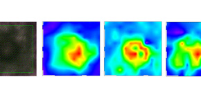
The experiment showed that Lewy bodies in the brain of Parkinson's disease patients had many β sheet structures*1. This supports the validity of in-vitro studies in the past. It was also found that the rate of β sheet structures was higher in the halo of a Lewy body than in the core of a Lewy body and the core was lipid-rich. These findings will lead to the elucidation of the mystery of the formation of Lewy bodies.
This research was featured in the electronic version of Scientific Reports on Tuesday, December 1, 2015.
Abstract
LBs, which mainly consist of α-synuclein (α-syn), are neuropathological hallmarks of patients with PD. The fine structure of LBs is unknown, and LBs cannot be made artificially. Nevertheless, many studies have described fibrillisation using recombinant α-syn purified from E. coli. An extremely fundamental problem is whether the structure of LBs is the same as that of recombinant amyloid fibrils. Thus, we used synchrotron FTIRM to analyse the fine structure of LBs in the brain of PD patients. Our results showed a shift in the infrared spectrum that indicates abundance of a β-sheet-rich structure in LBs. Also, 2D infrared mapping of LBs revealed that the content of the β-sheet structure is higher in the peripheral region (halo) than in the core, and the central region (core) contains a large amount of proteins and lipids.

The analysis results for a typical Lewy bodies
The central region (core) contains a large amount of proteins (second from left) and lipids (fourth from left). In contrast, the content of the β-sheet structure (third from left) is higher in the peripheral region (halo) than in the core.
To learn more about this research, please view the full research report entitled " Synchrotron FTIR micro-spectroscopy for structural analysis of Lewy bodies in the brain of Parkinson’s disease patients " at this page of the Scientific Reports website.
Related links