
Successful analysis of nanoparticle size distribution and structure differentiation using focused hard XFEL pulses
Opens up new frontier!
A group of researchers under the combined leadership of Yukio TAKAHASHI (Department of Precision Science and Technology, Graduate School of Engineering, Osaka University; RIKEN SPring-8 Center), Masayoshi NAKASAKO (Dept. of Physics, School of Engineering, Keio University), and Masaki YAMAMOTO (RIKEN SPring-8 Center) demonstrated success in the coherent diffraction imaging analysis of nanoparticles regarding size distribution and individual projection structures using focused hard X-ray free-electron laser pulses. X-ray free-electron lasers (XFELs) are a promising new source of X-ray imaging analysis. They made use of the SPring-8 Angstrom Compact Free Electron Laser (SACLA) in Japan for their research, one of two locations according to their research report for XFELs in the world.

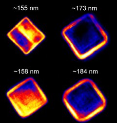
At SACLA they conducted the first demonstration of imaging analysis based on CXDI (coherent X-ray diffraction imaging, a lensless X-ray imaging technique) with focused hard XFEL pulses at SACLA. This CXDI using XFEL pulses was conducted on nanoparticles made from the metals silver and gold, "cubes" of silver and "boxes" of silver and gold. The methods they employed enabled them to reconstruct the two-dimensional electron density projection images with outstanding sub-10 nm resolution and the simultaneous analysis of the size distribution of nanoparticles of the silver cubes and gold/silver boxes and their statistical properties. It is their opinion that "coherent diffraction imaging analysis with XFELs will open up a new frontier in the imaging of heterogeneous systems such as cells, aerosols, and nanomaterials."
Abstract
We report the first demonstration of the coherent diffraction imaging analysis of nanoparticles using focused hard X-ray free-electron laser pulses, allowing us to analyze the size distribution of particles as well as the electron density projection of individual particles. We measured 1000 single-shot coherent X-ray diffraction patterns of shape-controlled Ag nanocubes and Au/Ag nanoboxes and estimated the edge length from the speckle size of the coherent diffraction patterns. We then reconstructed the two-dimensional electron density projection with sub-10 nm resolution from selected coherent diffraction patterns. This method enables the simultaneous analysis of the size distribution of synthesized nanoparticles and the structures of particles at nanoscale resolution to address correlations between individual structures of components and the statistical properties in heterogeneous systems such as nanoparticles and cells.
To learn more about this research, please read the full research report entitled " Coherent Diffraction Imaging Analysis of Shape-Controlled Nanoparticles with Focused Hard X-ray Free-Electron Laser Pulses " at this page of the Nano Letters website.