Ptychographic-X-ray absorption fine structure (XAFS) technique visualizes oxygen diffusion of material with oxygen storage/release capability
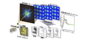
A group of researchers from RIKEN Spring-8 Center, Osaka University Graduate School of Engineering, and Nagoya University Research Center for Materials Science, developed a hard X-ray spectro-ptychography (ptychographic-X-ray absorption fine structure (XAFS)) technique to obtain X-ray absorption fine structure (XAFS) by using X-ray ptychography, visualizing oxygen diffusion of a material with oxygen storage/release capability.
X-ray ptychography, a coherent X-ray diffractive imaging technique, allows one to observe extended objects with a high-spatial resolution. Unlike conventional lens-based X-ray microscopy, X-ray ptychography reconstructs sample images from diffraction patterns by phase retrieval calculation. Thus, this technique can drastically improve the spatial resolution with X-ray microscopy that depended on the ability to manufacture lenses.
Meanwhile, XAFS is a technique for investigating the local structure and electronic state (valence and symmetry) of an X-ray absorption atom. XAFS spectroscopy is most frequently performed using synchrotron radiation. The micro-XAFS method is operated using microbeam X-ray to investigate a microscopic region in a sample, but the spatial resolution available on the device used to focus light is only about 100 nm, so the improvement of spatial resolution has been a challenge.
This joint group developed a ptychography-XAFS technique, a technique to obtain X-ray absorption fine structure (XAFS) using X-ray ptychography. They used platinum-supported cerium–zirconium oxide Pt/Ce 2 Zr 2 O x , as a sample. Platinum-supported cerium–zirconium oxide has an excellent oxygen storage/release capability and plays a role in controlling the amount of oxygen in the catalyst by supporting platinum, a major catalyst, in an exhaust gas purification catalyst system.
They performed hard X-ray ptychographic-XAFS measurements of Pt-supported Ce 2 Zr 2 O x particles using X-rays at SPring-8, the large synchrotron radiation facility, obtaining X-ray absorption fine structure (XAFS) of Pt/Ce 2 Zr 2 O x at a spatial resolution that surpasses those of the conventional micro XAFS methods. They also obtained two-dimensional mappings of the density and valence of Pt/Ce 2 Zr 2 O x by analyzing XAFS, and visualized oxygen diffusion.
This ptychography-XAFS will be applied to analysis of the nano-structures and chemical state of a variety of advanced functional materials in the future. Currently, it takes a couple of hours to measure a sample and its spatial resolution is the order of several tens of nanometers. However, at next-generation large synchrotron radiation facilities, in which x-ray energy and brightness will be 10-100 times greater than those available at Spring-8, the spatial resolution of a few nanometers can be achieved, promoting the design and development of advanced functional materials.
Abstract
The cerium density and valence in micrometer-size platinum-supported cerium–zirconium oxide Pt/Ce 2 Zr 2 O x (x=7–8) three-way catalyst particles were successfully mapped by hard X-ray spectro-ptychography (ptychographic-X-ray absorption fine structure, XAFS). The analysis of correlation between the Ce density and valence in ptychographic-XAFS images suggested the existence of several oxidation behaviors in the oxygen storage process in the Ce 2 Zr 2 O x particles. Ptychographic-XAFS will open up the nanoscale chemical imaging and structural analysis of heterogeneous catalysts.
Figure 1
Figure 2
Figure 3
To learn more about this research, please view the full research report entitled " Visualization of Heterogeneous Oxygen Storage Behavior in Platinum-Supported Cerium-Zirconium Oxide Three-Way Catalyst Particles by Hard X-ray Spectro-Ptychography " at this page of Angewandte Chemie International Edition .
Related links
