
Successful high resolution X-ray imaging of thick objects
Has excellent potential the enable three-dimensional high-resolution imaging of thick specimens
Under the leadership of TAKAHASHI Yukio , (Associate Professor, Graduate School of Engineering, Osaka University) SUZUKI Akihiro (doctoral candidate, fellow, Japan Society for the Promotion of Science) and ISHIKAWA Tetsuya (Director, SPring-8 Center, Riken) a group of researchers demonstrated that high resolution X-ray imaging of thick objects was indeed possible.
The ability of x-rays to penetrate even thick objects makes it possible to view the inside of thick objects and, at least in principle, it should be possible to make a high resolution microscope using x-rays as a source of illumination. However, it's not easy to observe the inside of thick objects at high resolution using x-rays. In x-ray imaging, projection approximation is used for image reconstruction; however, x-rays have wave-like properties so they spread out in a specimen rendering high resolution x-ray imaging of thick objects employing projection approximation unworkable.
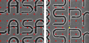
This group demonstrated high resolution x-ray ptychography using a multislice approach at the synchrotron radiation facility, SPring-8. When they measured a specimen 105 μm thick, they obtained some 50nm resolution, which was far higher than 192nm, the upper limit available under projection approximation.
Multislice x-ray ptychography has enabled researchers to observe nano structures and tissues in thick specimens, thus it will be used for non-destructive, high resolution, three-dimensional observation of micro wiring on 3D IC and bone structures of living organisms.
Abstract
We report the first demonstration of hard x-ray ptychography using a multislice approach, which can solve the problem of the limited spatial resolution under the projection approximation. We measured ptychographic diffraction patterns of a two-layered object with a 105 μm gap using 7 keV focused coherent x rays. We successfully reconstructed the phase map of each layer at ∼50 nm resolution using a multislice approach, while the resolution was worse than ∼192 nm under the projection approximation. The present method has the potential to enable the three-dimensional high-resolution observation of extended thick specimens in materials science and biology.

Figure 1

Figure 2

Figure 3
To learn more about this research, please read the full research report entitled " High-Resolution Multislice X-Ray Ptychography of Extended Thick Objects " at this page of the Physical Review Letters website.
Related link :