
Detailed Structure of the Sweat Gland Revealed
Researchers at Osaka University have characterized the structure of human sweat glands down to the single cell level; the findings clarify the functional components of these glands and their interactions with the vasculature and nervous system, and thus could lead to treatments for sweating disorders
The basic structure of human sweat glands consists of a coiled secretory region deep in the skin and then a tube or duct that conveys the sweat to a pore at the skin surface. However, it has been difficult to obtain more detailed structural insights due to technical limitations and the structural complexity of these glands.
In a major breakthrough that boosts our understanding of how we sweat, a team at Osaka University has applied a recently developed technique called whole-mount immunostaining to study sweat glands, revealing their structure down to the single-cell level and thus shedding light on the functions of their different components. The obtained findings should lead to treatments for disorders of the perspiratory system.
Conventional approaches for studying structural features of tissues involve cutting blocks of tissue into thin sections, and then viewing these sections after applying certain stains or dyes to resolve the features. Although these sections can then be reconstructed in a series to approximate the 3D arrangement of the tissue, this has a drawback in that the sectioning and preparation procedures can disrupt minute structural features. As reported in the journal PLOS One, the Osaka University team has now overcome these issues by applying whole-mount immunostaining to sweat glands after removing adjacent connective tissue in the skin, enabling anatomic features to be observed in a completely intact state.
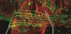
“Whole-mount immunostaining allows us to view the sweat gland structures seamlessly, without interruptions for sectioning, which is very important given that part of the gland has tubes that are entangled with each other in a very complicated way,” Kiyotoshi Sekiguchi says. “We revealed the different cross-sectional shapes of these tubes and the cells that they’re made of.”
The team’s findings reveal the importance of cells called myoepithelial cells working in unison to induce contraction of the secretory part of sweat glands to convey sweat to the exterior. The results also show how nerves and blood vessels interact with the various components of sweat glands, providing hints about how regulation of sweating could go awry.
“This knowledge about sweat gland structure and function could have many clinical applications, and even lead to strategies for treating heatstroke,” Ryuichiro Kurata adds. “Because of the high-resolution findings that our approach provides, we can even determine the density of nerve fibers in sweat glands, helping to diagnose specific sweating-related disorders and to select appropriate treatments.”
Abstract
Because sweat secretion is facilitated by mechanical contraction of sweat gland structures, understanding their structure-function relationship could lead to more effective treatments for patients with sweat gland disorders such as heat stroke. Conventional histological studies have shown that sweat glands are three-dimensionally coiled tubular structures consisting of ducts and secretory portions, although their detailed structural anatomy remains unclear. To better understand the details of the three-dimensional (3D) coiled structures of sweat glands, a whole-mount staining method was employed to visualize 3D coiled gland structures with sweat gland markers for ductal luminal, ductal basal, secretory luminal, and myoepithelial cells. Imaging the 3D coiled gland structures demonstrated that the ducts and secretory portions were comprised of distinct tubular structures. Ductal tubules were occasionally bent, while secretory tubules were frequently bent and formed a self-entangled coiled structure. Whole-mount staining of complex coiled gland structures also revealed the detailed 3D cellular arrangements in the individual sweat gland compartments. Ducts were composed of regularly arranged cuboidal shaped cells, while secretory portions were surrounded by myoepithelial cells longitudinally elongated along entangled secretory tubules. Whole-mount staining was also used to visualize the spatial arrangement of blood vessels and nerve fibers, both of which facilitate sweat secretion. The blood vessels ran longitudinally parallel to the sweat gland tubules, while nerve fibers wrapped around secretory tubules, but not ductal tubules. Taken together, whole-mount staining of sweat glands revealed the 3D cell shapes and arrangements of complex coiled gland structures and provides insights into the mechanical contraction of coiled gland structures during sweat secretion.
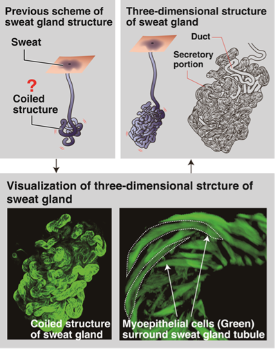
Figure 1. Detail three-dimensional structure of human sweat gland
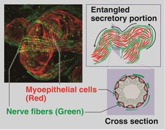
Figure 2. Three-dimensional arrangements of myoepithelial cells and nerve fibers at secretory portion of human sweat glands.
To learn more about this research, please view the full research report entitled " Three-dimensional Cell Shapes and Arrangements in Human Sweat Glands as Revealed by Whole-mount Immunostaining " at this page of the PLOS ONE website.
Related links
- Laboratory of Advanced Cosmetic Science, Graduate School of Pharmaceutical Sciences, Osaka University
- Division of Matrixome Research and Application Institute for Protein Research, Osaka University (link in Japanese)
- Department of Dermatology Course of Integrated Medicine, Graduate School of Medicine, Osaka University