
Molecule Causing Obesity-associated Chronic Inflammation Identified
A breakthrough discovery in breaking the link between obesity and lifestyle-related diseases
A group of researchers led by MAEDA Norikazu (Assistant Professor, Department of Metabolic Medicine) and ISHII Masaru (Professor, Immunology) at the Graduate School of Medicine, Osaka University, examined in detail the process of chronic inflammation associated with obesity by using their bio-imaging technique and found a molecule causing obesity-associated chronic inflammation.
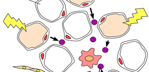
Usually, eating high calorie meals for a couple of weeks will result in obesity and fat cells becoming enlarged. By making use of bio-imaging techniques, this group found that just 5 days after taking high calorie meals, during the period before becoming obese, inflammatory macrophages in fat tissues became active. During this period, there were no visible changes in fat cells; however, their nature was changing. They released S100A8, a factor to activate migration and inflammation of macrophages.
This group succeeded in preventing macrophages from migrating and chronic inflammation associated with obesity from progressing by suppressing 100A8. It is highly expected that suppressing 100A8 will lead to the development of epoch-making therapeutic methods for preventing lifestyle-related diseases associated with chronic inflammation.
Abstract
Chronic low-grade inflammation of adipose tissue plays a crucial role in the pathophysiology of obesity. Immunohistological microscopic analysis in obese fat tissue has demonstrated the infiltration of several immune cells such as macrophages, but dynamics of immune cells have not been fully elucidated and clarified. Here, by using intravital multiphoton imaging technique, to our knowledge for the first time, we analyzed and visualized the inflammatory processes in adipose tissue under high-fat and high-sucrose (HF/HS) diet with lysozyme M-EGFP transgenic (LysM EGFP ) mice whose EGFP was specifically expressed in the myelomonocytic lineage. Mobility of LysM EGFP -positive macrophages was shown to be activated just 5 d after HF/HS diet, when the distinct hypertrophy of adipocytes and the accumulation of macrophages still have not become prominent. Significant increase of S100A8 was detected in mature adipocyte fraction just 5 d after HF/HS diet. Recombinant S100A8 protein stimulated chemotactic migration in vitro and in vivo, as well as induced proinflammatory molecules, both macrophages and adipocytes, such as TNF-α and chemokine (C-C motif) ligand 2. Finally,an antibody against S100A8 efficiently suppressed the HF/HS diet-induced initial inflammatory change, i.e., increased mobilization of adipose LysM EGFP -positive macrophages, and ameliorated HF/HS diet-induced insulin resistance. In conclusion, time-lapse intravital multiphoton imaging of adipose tissues identified the very early event exhibiting increased mobility of macrophages, which may be triggered by increased expression of adipose S100A8 and results in progression of chronic inflammation in situ.
A significant increase of S100A8 was detected in fat tissue. S100A8 stimulated chemotactic migration.
To learn more about this research, please view the full research report entitled " Visualized macrophage dynamics and significance of S100A8 in obese fat " at this page of the Proceedings of the National Academy of Science of the USA (PNAS) website.
Related link