
Formation site of autophagosomal membranes identified
Under the leadership of HAMASAKI Maho , Assistant Professor and YOSHIMORI Tamotsu , Professor, Graduate School of Frontier Biosciences, Graduate School of Medicine, Osaka University and FURUTA Nobumichi , Assistant Professor and AMANO Atsuo , Professor, Graduate School of Dentistry, Osaka University, a group of researchers clarified the location from which autophagosomal membranes originate in the formation of double membrane-bound autophagosomes in mammalian cells.
Autophagy is a body mechanism that assists the body in preventing diseases such as Alzheimer's disease, cancer, and heart failure from developing by degrading and recycling internal portions of cells. In autophagy, autophagosomes are formed in cells and degrade and recycle old and damaged proteins and organelles. The origin of autophagosomal membranes has remained unclear, but this group has clarified the location and mechanism behind their formation.
The identification of the location and mechanism site of autophagosomal membranes will help researchers in the development of treatments that can make use of regulating the formation of autophagosomes.
Abstract
Autophagy is a tightly regulated intracellular bulk degradation/recycling system that has fundamental roles in cellular homeostasis1. Autophagy is initiated by isolation membranes, which form and elongate as they engulf portions of the cytoplasm and organelles. Eventually isolation membranes close to form double membrane-bound autophagosomes and fuse with lysosomes to degrade their contents. The physiological role of autophagy has been determined since its discovery, but the origin of autophagosomal membranes has remained unclear. At present, there is much controversy about the organelle from which the membranes originate—the endoplasmic reticulum (ER), mitochondria and plasma membrane1, 2. Here we show that autophagosomes form at the ER–mitochondria contact site in mammalian cells. Imaging data reveal that the pre-autophagosome/autophagosome marker ATG14 (also known as ATG14L) relocalizes to the ER–mitochondria contact site after starvation, and the autophagosome-formation marker ATG5 also localizes at the site until formation is complete. Subcellular fractionation showed that ATG14 co-fractionates in the mitochondria-associated ER membrane3, 4, 5 fraction under starvation conditions. Disruption of the ER–mitochondria contact site prevents the formation of ATG14 puncta. The ER-resident SNARE protein syntaxin 17 (STX17) binds ATG14 and recruits it to the ER–mitochondria contact site. These results provide new insight into organelle biogenesis by demonstrating that the ER–mitochondria contact site is important in autophagosome formation.

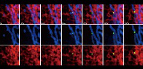
Figure 1

Figure 2
To learn more about this research, please read the full research report entitled " Autophagosomes form at ER–mitochondria contact sites " at this page of the Nature website.
Related link :