
World’s lightest, palm-sized gamma-ray camera for medical purposes
Multi-angle shooting and multi-color 3D imaging of a live mouse achieved
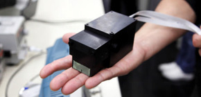
A group of researchers from Waseda University, in cooperation with Osaka University, National Institutes for Quantum and Radiological Science and Technology, and Hamamatsu Photonics K.K., invented the world’s lightest camera (580g) capable of visualizing gamma rays. With this camera, this group succeeded in simultaneous 3D in vivo imaging of a mouse administered with three different radioactive tracers, a world first.
Radiographs typified by X-ray images provide only monochrome images without energy information. Also, in positron emission tomography (PET) used for early detection of cancer and Alzheimer’s disease, 511 keV gamma rays are detected and PET images are monochrome.
Thus, if gamma rays of arbitrary energies could be easily visualized, it will become possible to simultaneously trace multiple markers with different properties and positions where radioactive tracers concentrate. One could expect that the amount of available information would increase as drastically as replacing a black and white TV with a color one.
With this thought, this group improved the performance of a Compton camera for environmental measurements, developing the world’s lightest medical Compton camera with a high spatial resolution. Using this camera, this group administered radioactive tracers that emit different gamma rays to a living mouse, demonstrating that iodine (131I, 364keV) was taken into the thyroid, strontium (85Sr, 514keV) into bones, zinc (65Zn, 1116keV) into the liver, lungs, heart, and pancreas in the short time of 2 hours at a high resolution of about 3mm. Downsizing the detector with high resolution as much as possible, this group achieved filming from multiple angles, obtaining 3D color images with uniform image intensity.
Moving forward, it is anticipated that this group’s achievement will lead to the development of gamma ray cameras closer to the vision of a human eye and extend the possibilities of next-generation molecular imaging.
Abstract
In the field of nuclear medicine, single photon emission tomography and positron emission tomography are the two most common techniques in molecular imaging, but the available radioactive tracers have been limited either by energy range or difficulties in production and delivery. Thus, the use of a Compton camera, which features gamma-ray imaging of arbitrary energies from a few hundred keV to more than MeV, is eagerly awaited along with potential new tracers which have never been used in current modalities. In this paper, we developed an ultra-compact Compton camera that weighs only 580 g. The camera consists of fine-pixelized Ce-doped Gd 3 Al 2 Ga 3 O 12 scintillators coupled with multi-pixel photon counter arrays. We first investigated the 3-D imaging capability of our camera system for a diffuse source of a planar geometry, and then conducted small animal imaging as pre-clinical evaluation. For the first time, we successfully carried out the 3-D color imaging of a live mouse in just 2 h. By using tri-color gamma-ray fusion images, we confirmed that 131 I, 85 Sr, and 65 Zn can be new tracers that concentrate in each target organ.

Figure 1

Figure 2

Figure 3
To learn more about this research, please view the full research report entitled " First demonstration of multi-color 3-D in vivo imaging using ultra-compact Compton camera " at this page of the Scientific Reports website.
Related links