
Successful determination of crystal structures by using sulfur atoms of proteins at SACLA
Will enable analysis of various unknown protein structures, or potential drug targets
Michihiro Sugahara (Researcher, SPring-8 Center, RIKEN)
IWATA So (Group Director; Professor, Graduate School of Medicine, Kyoto University)
YABASHI Makina (Group Director, Beamline Construction Team, XFEL Project Head Office)
NAKANE Tomonori (Researcher, Graduate School of Science, The University of Tokyo)
SUZUKI Mamoru (Associate Professor, Research Center for State-of-the-Art Functional Protein Analysis, Institute for Protein Research, Osaka University)
TONO Kensuke (Team Leader, XFEL Project Head Office, Japan Synchrotron Radiation Research Institute (JASRI))
A group of researchers succeeded in determining crystal structures by using sulfur atoms of proteins. Using the method of Serial Femtosecond Crystallography (SFX), they achieved this with the x-ray laser at the X-ray Free Electron Laser facility, SPring-8 Angstrom Compact Free Electron Laser (SACLA). In SFX, using the x-ray laser at SACLA, the structures of minute crystal proteins of μm or less were made available without causing radiation damage, which had been a challenge.
Generally, in X-ray crystallography, protein models with similar structure are used for determining protein structures. However, if there are no proteins with similar structures, the structure is determined by using the anomalous dispersion effect from platinum- and mercury-containing heavy-atom derivatives of protein crystals.
In recent years, in order to save the effort of heavy-atom derivatization, a sulfur single wavelength anomalous dispersion (SSAD) employing sulfur-containing amino acids has been used. However, even through the use of the x-ray at SPring-8, it is hard to determine protein structures by using SSAD when the protein crystal size is not big enough and/or the crystal suffers from radiation damage.
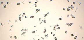
This joint group succeeded in determining structures by using sulfur atoms of lysozyme proteins with a grease matrix method that they had previously developed, which enabled them to perform diffraction of various proteins in a small amount of sample.
With SSAD using SACLA, it will become possible to determine structures of proteins, even those with insufficient crystal size and/or radiation-damage problems. It is also expected that this technique will be used for structural analysis of proteins, or potential drug targets.
Abstract
Serial femtosecond crystallography (SFX) allows structures to be determined with minimal radiation damage. However, phasing native crystals in SFX is not very common. Here, the structure determination of native lysozyme from single-wavelength anomalous diffraction (SAD) by utilizing the anomalous signal of sulfur and chlorine at a wavelength of 1.77 Å is successfully demonstrated. This sulfur SAD method can be applied to a wide range of proteins, which will improve the determination of native crystal structures.

Figure 1

Figure 2
To learn more about this research, please view the full research report entitled " Native sulfur/chlorine SAD phasing for serial femtosecond crystallography " at this page of the Acta Crystallographica Section D: Biological Crystallography website.
Related links
- Press release from RIKEN (November 11, 2014) (link in Japanese)