
Photoproteins Visible to the Naked Eye Developed
Activity of pluripotent cells quanitifiable in real time
A group of researchers led by NAGAI Takeharu (Professor, Institute of Scientific and Industrial Research, Osaka University) and OKADA Yasushi (Team leader, Quantitative Biology Center, RIKEN) succeeded in developing two types of super-high luminosity photoproteins - cyan and orange - that shine more brightly than the conventional, lime green super-high luminosity photoprotein developed in 2012, the Nano-lantern. Since these new photoproteins are 20 times brighter than conventional photoproteins, one can observe emission of light without using a special highly sensitive camera.
Fluorescence live imaging has become an essential methodology in modern cell biology. However, fluorescence requires excitation light, which can sometimes cause potential problems, such as autofluorescence, phototoxicity, and photobleaching. Furthermore, combined with recent optogenetic tools, the light illumination can trigger their unintended activation. Because luminescence imaging does not require excitation light, it is a good candidate as an alternative imaging modality to circumvent these problems. The application of luminescence imaging, however, has been limited by the two drawbacks of existing luminescent protein probes, such as luciferases: namely, low brightness and poor color variants. Here, we report the development of bright cyan and orange luminescent proteins by extending our previous development of the bright yellowish-green luminescent protein Nano-lantern. The color change and the enhancement of brightness were both achieved by bioluminescence resonance energy transfer (BRET) from enhanced Renilla luciferase to a fluorescent protein. The brightness of these cyan and orange Nano-lanterns was ∼20 times brighter than wild-type Renilla luciferase, which allowed us to perform multicolor live imaging of intracellular submicron structures. The rapid dynamics of endosomes and peroxisomes were visualized at around 1-s temporal resolution, and the slow dynamics of focal adhesions were continuously imaged for longer than a few hours without photobleaching or photodamage. In addition, we extended the application of these multicolor Nano-lanterns to simultaneous monitoring of multiple gene expression or Ca2+ dynamics in different cellular compartments in a single cell.

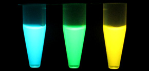
Left: Luminescence of 3-color recombinant Nano-lantern proteins. Right: Inhomogeneous expression of pluripotency markers in a single colony of ES cells. Luminescence signals of reporters for Oct4 (Cyan Nano-lantern), Nanog (Yellow Nano-lantern), and Sox2 (Orange Nano-lantern) were merged and overlaid with the bright-field image. (Scale bar: 100 μm.)
To learn more about this research, please view the full research report entitled " Expanded palette of Nano-lanterns for real-time
multicolor luminescence imaging " at this page of the Proceedings of the National Academy of Sciences of the United States of America website.
Related Link