
Bright luminescent proteins capable of detecting single molecules developed
New sensitive measurement technology of life phenomena
Bio-imaging, a technology for visualizing living things, has developed significantly, but most subjects of bio-imaging are present in small amounts in cells, so autofluorescence hides their signals, which often makes fluorescent observation difficult. In addition, irradiation of strong light is necessary for measuring high-speed life phenomena, which may disturb the physiological state of cells.
On the other hand, chemiluminescence is emission of light (luminescence) from oxidation catalysis reaction of a light-emitting compound luciferin by luciferase, an enzyme protein (luminescent protein). This technology is promising as a next-generation imaging technology that can overcome fluorescent proteins’ problems because it does not need exciting light irradiation. However, luminescent proteins with high intensity of light and multiple color variants have not been reported, so it was difficult to perform simultaneous imaging of multiple life phenomena and protein dynamics.
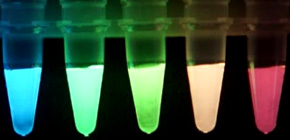
A group of researchers led by Professor NAGAI Takeharu at The Institute of Scientific and Industrial Research succeeded in developing 5-color luminescent proteins by improving the luminescent protein Nano-lantern that they developed in 2012 and 2015. This group developed an enhanced Nano-lantern (eNL), a protein which has a brightness of 2 to 10 times that of conventional ones and produces light of cyan, green, greenish yellow, orange, and red by concatenation of luminescent proteins with high enzymatic activity with 5 color hues of fluorescent proteins. This has allowed the group to perform measurements of 5 fine structures in a cell at the same time. Furthermore, this group succeeded in detecting binding and diffusing of a single protein molecule by chemiluminescence through the use of eNL.
This type of measurement has been performed using fluorescent proteins while irradiating light of a few times the intensity of sunlight in summer, so autofluorescence and phototoxicity have been a problem. However, eNL does not need exciting light irradiation from outside, so there is no autofluorescence or phototoxicity. Furthermore, this group also developed a chemiluminescence sensor, which can detect calcium ions in a cell by improving eNL. With this, this group succeeded in measuring Ca 2+ dynamics through imaging of cardiac muscle cells derived from iPS cells at a high speed of 60 frames per second.
These chemiluminescence sensors will make it possible to measure cells in a biological state. It is hoped that they will make great contributions to research in the fields of medicine and pharmacodynamics. Using this group’s achievements, simultaneous observation of multiple states of minute amounts of proteins under biological conditions can be performed, which will contribute to the clarification of protein networks. In addition, a red variant of eNL, which has high intensity of light and excellent tissue permeability, enables researchers to observe signals inside the body from outside in real time at high sensitivity, making it possible to observe cancer stem cells from outside the body. This will lead to the clarification of causes of many diseases and achievement of more effective screening for drug development.
Abstract
Luminescence imaging has gained attention as a promising bio-imaging modality in situations where fluorescence imaging cannot be applied. However, wider application to multicolour and dynamic imaging is limited by the lack of bright luminescent proteins with emissions across the visible spectrum. Here we report five new spectral variants of the bright luminescent protein, enhanced Nano-lantern (eNL), made by concatenation of the brightest luciferase, NanoLuc, with various colour hues of fluorescent proteins. eNLs allow five-colour live-cell imaging, as well as detection of single protein complexes and even single molecules. We also develop an eNL-based Ca 2+ indicator with a 500% signal change, which can image spontaneous Ca 2+ dynamics in cardiomyocyte and neural cell models. These eNL probes facilitate not only multicolour imaging in living cells but also sensitive imaging of a wide repertoire of proteins, even at very low expression levels.

Figure 1

Figure 2
To learn more about this research, please view the full research report entitled “ Five color variants of bright luminescent protein for real-time multicolor bioimaging ” at this page of the Nature Communications website.
Related link