
Dynamic visualization of osteoclast function
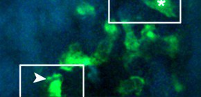
Under the leadership of ISHII Masaru , Professor, Immunology Frontier Research Center (IFReC), Osaka University, a group of researchers succeeded in visualizing live osteoclasts resorption for the first time in the world. This visualization was made possible by using intravital multiphoton microscopy and clarified that there were two types of osteoclasts -- moving-nonresorptive (N) osteoclasts and static-resorptive (R) ones. Furthermore, they discovered that osteoclast-activation factor RANKL significantly shortened the time necessary for N-to-R conversion. They also clarified that in conditions such as in bone-resorptive inflammatory diseases, not only the total number of osteoclasts, but also the number of R osteoclasts increased. Such findings indicate that prospects for resorption-switching may represent a therapeutic technique for certain conditions.
Abstract
Osteoclasts are bone resorbing, multinucleate cells that differentiate from mononuclear macrophage/monocyte-lineage hematopoietic precursor cells. Although previous studies have revealed important molecular signals, how the bone resorptive functions of such cells are controlled in vivo remains less well characterized. Here, we visualized fluorescently labeled mature osteoclasts in intact mouse bone tissues using intravital multiphoton microscopy. Within this mature population, we observed cells with distinct motility behaviors and function, with the relative proportion of static – bone resorptive (R) to moving – nonresorptive (N) varying in accordance with the pathophysiological conditions of the bone. We also found that rapid application of the osteoclast-activation factor RANKL converted many N osteoclasts to R, suggesting a novel point of action in RANKL-mediated control of mature osteoclast function. Furthermore, we showed that Th17 cells, a subset of RANKL-expressing CD4+ T cells, could induce rapid N-to-R conversion of mature osteoclasts via cell-cell contact. These findings provide new insights into the activities of mature osteoclasts in situ and identify actions of RANKL-expressing Th17 cells in inflammatory bone destruction.

Figure 1
To learn more about this research, please read the full research report entitled " Dynamic visualization of RANKL and Th17-mediated osteoclast function " at this page of The Journal of Clinical Investigation website.
Related links :