
Intravital imaging of bone dissolving cell function achieved
Observation of osteoclasts made possible
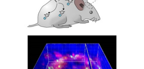
A group of researchers led by Professor KIKUCHI Kazuya and Professor ISHII Masaru at the Immunology Frontier Research Center, Osaka University produced fluorescent probes for visualizing a site where osteoclasts were actually dissolving the bone, succeeding in evaluation of the function of osteoclasts in vivo with their own intravital imaging through two-photon excitation microscopy (TPEM).
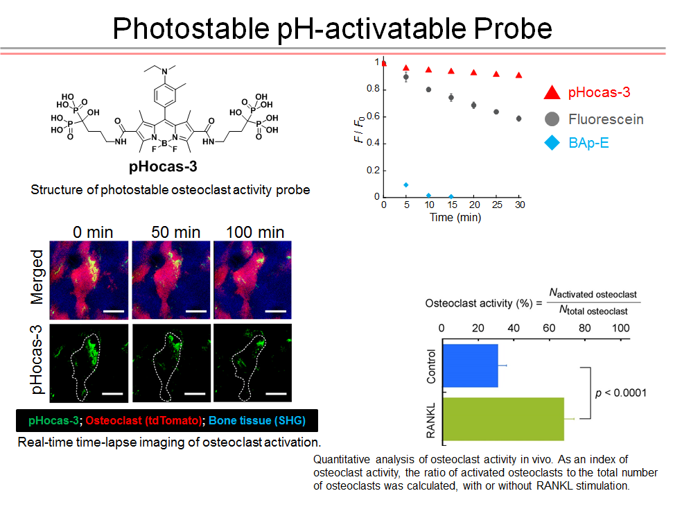
This group demonstrated that the selective visualization of osteoclast activity became possible by letting small molecular probes (SMPs) have (1) the function of selectively delivering to certain tissues where target cells were present and (2) a functional switch for emitting fluorescence at the site of bone resorption.
Furthermore, this group succeeded in real-time imaging of local change and activity change of cells as well as quantitating the intensity of bone resorption by labeling target cells with fluorescent proteins and by simultaneously detecting SMPs and fluorescent signals from fluorescent proteins.
This group established an intravital imaging method for performing easy and stable measurements in order to detect functioning osteoclasts. It is notable that this group achieved imaging of mice only by subcutaneously injecting molecular probes into mice through the optimization of molecular delivery.
As there were no molecular probes to be used for intravital imaging, this group’s achievement will have a great impact on the field of in vivo imaging. With conventional methods using fluorescent proteins, it was possible to get information about osteoclast localization, but not about osteoclast activity. However, with this method, it’s possible to get information on osteoclast activity easily and quickly, so it is expected that this method will be useful in early diagnosis of an affected area and screening of new drugs, greatly contributing to the medical and industrial worlds.
This study is interdisciplinary research crossing over fields of study such as molecular design based on physical chemistry principles, the creation of functional fluorescent probes by synthetic organic chemistry, and the clarification of intravital mechanisms using immunological knowledge and technology, and will make contributions to fields related to medicine, chemistry, and measurement device manufactures. This study has social and academic significance in that basic research has developed into medical research application.
Abstract
Intravital imaging by two-photon excitation microscopy (TPEM) has been widely used to visualize cell functions. However, small molecular probes (SMPs), commonly used for cell imaging, cannot be simply applied to intravital imaging because of the challenge of delivering them into target tissues, as well as their undesirable physicochemical properties for TPEM imaging. Here, we designed and developed a functional SMP with an active-targeting moiety, higher photostability, and a fluorescence switch and then imaged target cell activity by injecting the SMP into living mice. The combination of the rationally designed SMP with a fluorescent protein as a reporter of cell localization enabled quantitation of osteoclast activity and time-lapse imaging of its in vivo function associated with changes in cell deformation and membrane fluctuations. Real-time imaging revealed heterogenic behaviors of osteoclasts in vivo and provided insights into the mechanism of bone resorption.

To learn more about this research, please view the full research report entitled “ Real-time intravital imaging of pH variation associated with osteoclast activity ” at this page of the Nature Chemical Biology website.
Related links
- World Premier International Research Center, Immunology Frontier Research Center, Osaka University (WPI-IFReC)
- Kikuchi Laboratory, Laboratory of Chemical Biology, Division of Advanced Science and Biotechnology, Department of Material and Life Science, Graduate School of Engineering, Osaka University
- Department of Immunology and Cell Biology, Graduate School of Medicine and Frontier Biosciences, Osaka University
- EurekAlert!
- AlphaGalileo
- BioOptics World