
Development of new method for utilization of coherent x-rays
Generation of x-ray vortex beams by visualizing dislocation strain fields in materials
Under the leadership of TAKAHASHI Yukio , Associate Professor, Graduate School of Engineering, Osaka University, and ISHIKAWA Tetsuya , Director, SPring-8 Center, Harima Institute, Riken, a group of researchers have developed a new method for the utilization of coherent x-rays, generating x-ray vortex beams by visualizing dislocation strain fields in materials.
Dislocations are line defects in a crystal lattice and have strain fields arising from distortions at their cores. Strain fields strongly influence mechanical properties and electrical conductivity of materials. X-ray topography and transmission electron microscopy (TEM) are the two most common techniques used for studying dislocations, but they have limitations in spatial resolution of thick samples.
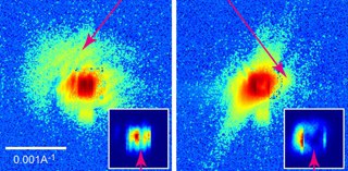
In the Beamline BL29XUL at the synchrotron radiation facility SPring-8, Department of Physical Sciences I, Riken, this group of researchers developed Bragg x-ray ptychography and succeeded in visualizing dislocation strain fields in a 1-μm thick silicon crystal at the nanometer-scale spatial resolution. The development of new structural materials and semiconductor materials using this visualization technology are expected.
This group also discovered that x-ray vortex beams with helical wavefront could be generated by using dislocation strain fields in silicon crystals. X-ray vortex beams can control orbital angular momentum (OAM) modes by selecting the dislocation singularity and diffraction index. The production of vortex beams using visible light and electron beams have been reported many times, but there were few reports on ones using x-rays. This group's achievements are really interesting from the viewpoint of X-ray optics. The presence of strong dichroic effects induced by x-ray beams is predicted. This group's results will be applied in analyzing complicated electronic structure in materials.
Abstract
We experimentally demonstrate the visualization of nanoscale dislocation strain fields in a thick silicon single crystal by a coherent diffraction imaging technique called Bragg x-ray ptychography. We also propose that the x-ray microbeam carrying orbital angular momentum is selectively produced by coherent Bragg diffraction from dislocation singularities in crystals. This work not only provides us with a tool for characterizing dislocation strain fields buried within extended crystals but also opens up new scientific opportunities in femtosecond spectroscopy using x-ray free-electron lasers.

Figure 1

Figure 2

Figure 3

Figure 4
To learn more about this research, please read the full research report entitled " Bragg x-ray ptychography of a silicon crystal: Visualization of the dislocation strain field and the production of a vortex beam " at this page of the Pysical Review B website.
Related links :