
Successful verification of a new technique, dark-field X-ray ptychography
A step towards the world’s best spatial resolution and sensitivity by X-ray imaging
X-ray ptychography, a coherent X-ray diffractive imaging technique, has high spatial resolution that is not available by conventional X-ray microscopes, so it has been actively studied as a next-generation X-ray imaging technology primarily at radiation facilities in recent years. In order to observe specimens at high spatial resolution and sensitivity through X-ray ptychography, a large dynamic range of intensities of diffraction patterns has to be measured. Therefore, a high-intensity coherent x-ray source and a high-performance two-dimensional X-ray detector are necessary.
Regarding the high-intensity coherent X-ray source, high-intensity low-emittance radiation sources are actively being developed all over the world, and the coherence of emitted light is improving. As for the high-performance two-dimensional X-ray detector, the emergence of hybrid pixel detectors with high photon-counting rates in two-dimensional X-ray detectors that are sensitized for high-energy X-rays has made it possible to obtain a dynamic range of diffraction intensity patterns in a short period of time. However, hybrid pixel detectors are expensive, and it is expected that the current photon-counting rate will become insufficient for use as a radiation source in the future.
A group of researchers led by Associate Professor TAKAHASHI Yukio at the Graduate School of Engineering, Osaka University and Team Leader at RIKEN, and Graduate Student SUZUKI Akihiro (currently Assistant Professor at the Research Institute for Electronic Science, Hokkaido University), succeeded in verifying dark-field X-ray ptychography.
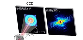
This group carried out the verification of a new method, dark-field X-ray ptychography, which compressed the dynamic range of diffraction intensity patterns to one-1,000th by combining in-line holography, which uses scattered X-rays from microstructures as reference beams, and dark-field X-ray ptychography at the large synchrotron radiation facility SPring-8, succeeding in high-resolution imaging of biological specimens.
This group’s achievement has made imaging with high spatial resolution and sensitivity by X-ray ptychography possible even when using an inexpensive two-dimensional X-ray detector with a small photon-counting rate, which will lead to the dissemination of this technology. Furthermore, the world’s highest spatial resolution and sensitivity will be available through the use of high-performance two-dimensional X-ray detectors.
Other countries have achieved spatial resolution and sensitivity equivalent to this group’s results by making full use of expensive hybrid pixel array detectors. This group achieved the world’s best spatial resolution and sensitivity relatively cheaply by using a charge-coupled device (CCD) detector with a small photon detection rate, so the dissemination of this technique will be facilitated. It is hoped that the world’s top spatial resolution and sensitivity will be available through the measurement of dark-field X-ray ptychography by using hybrid pixel array detectors.
Abstract
The phase shift of light or electrons in objects is now necessary for probing weak-phase objects such as unstained biological specimens. Optical microscopy (OM) and transmission electron microscopy (TEM) have been used to observe weak-phase objects. However, conventional OM has low spatial resolution and TEM is limited to thin specimens. Here, we report on the development of dark-field X-ray ptychography, which combines X-ray ptychography and X-ray in-line holography, to observe weak-phase objects with a phase resolution better than 0.01 rad, a spatial resolution better than 15 nm, and a field of view larger than 5 μm. We apply this method to the observation of both the outline and magnetosomes of the magnetotactic bacteria MO-1. Observation of thick samples with high resolution is expected to find broad applications in not only biology but also materials science.
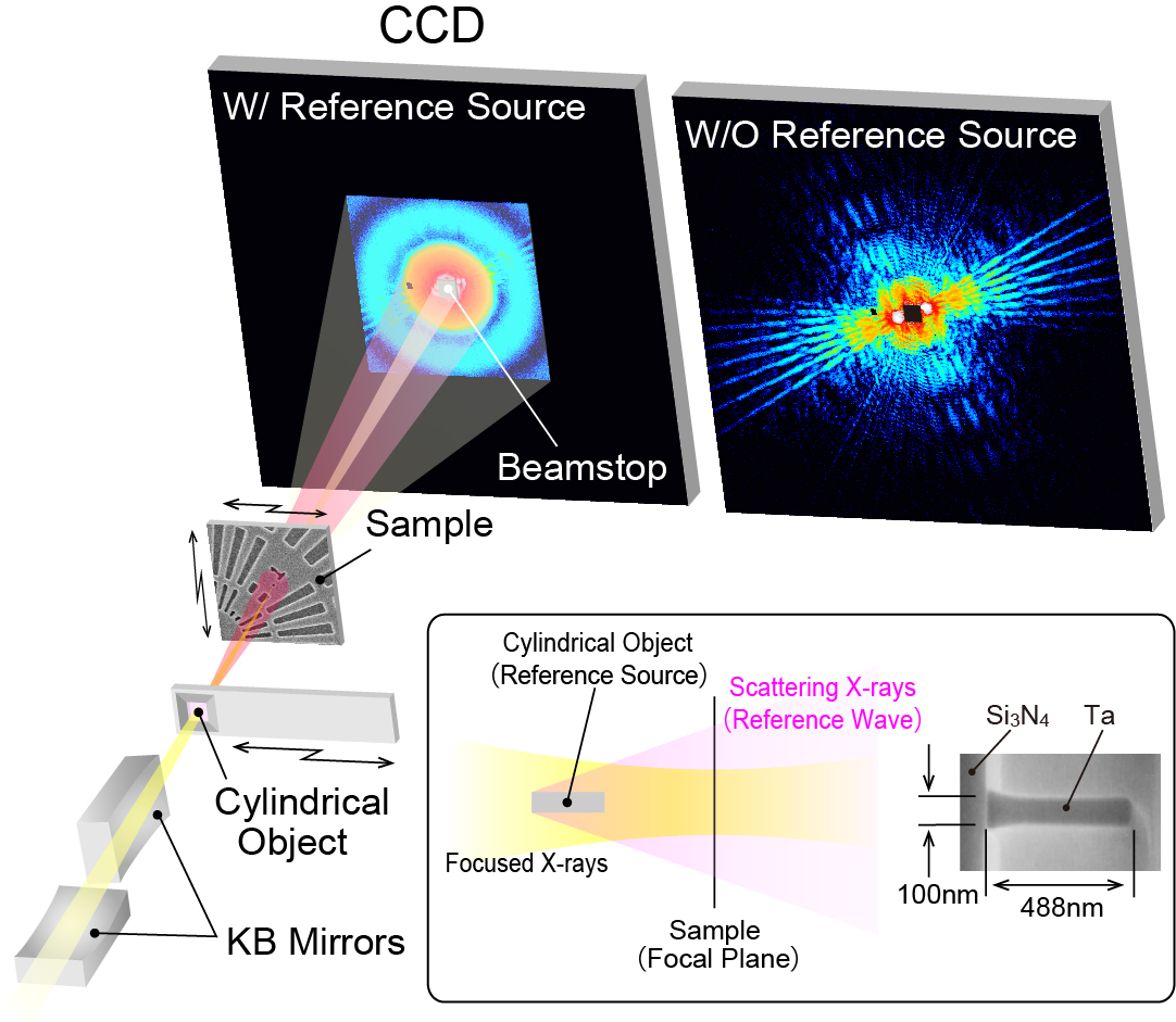
Fig. 1 Schematic view of experimental setup of dark-filed X-ray ptychography.
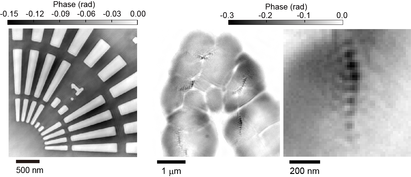
Fig. 2 (Left) Phase image of the test patterns obtained by dark-field X-ray ptychography. (Right) Phase image of MO-1 obtained by dark-field X-ray ptychography, and its enlarged image.
To learn more about this research, please view the full research report entitled “ Dark-field X-ray ptychography: Towards high-resolution imaging of thick and unstained biological specimens ” at this page of the Scientific Reports website.
Related link