Restoring a patient’s vision
Corneal epithelial cell sheet grafting and regeneration of the eye
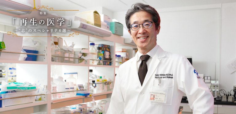
Professor Kohji Nishida Graduate School of Medicine, Osaka University
Professor Kohji Nishida was the first to prepare corneal epithelial cell sheets using iPS cells and graft them into patients with corneal diseases. He is also the leader in two-dimensionally reproducing the development of cells that constitute the eye using iPS cells. He has further made many globally acknowledged achievements, including the discovery of vascular endothelial stem cells, but says “I have never set a big goal. I am the type that makes small efforts day by day to solve problems that come up one at a time.”

Oral mucosa was where it started
Professor Nishida engaged in corneal grafting as an ophthalmologist. However, corneal grafting from another person has the problem of rejection and donors are difficult to find in Japan.
Therefore, he initiated research on regenerative medicine using the patients’ own cells around 2000. He selected the oral mucosa as the possible source of cells. Embryologically, the tissue of corneal epithelium closely resembles that of oral mucosa. In addition, a major advantage is that cells in the mouth are easy to harvest. If stem cells are collected from the patient’s mucosal tissue, cultured into sheets, and grafted, rejection can be avoided because the graft is the patient’s own tissue. In 2004, Professor Nishida reported the first transplantation of substitute corneal epithelium prepared from oral mucosa. The results of this procedure have been remarkable, including a case in which visual acuity of less than 0.01 recovered to 0.9. Presently, the physician-initiated clinical trial has been completed, and the study is approaching the stage in which manufacturing and marketing of sheets is expected to be approved.
However, it has also been learned that there are “limitations” with epithelial cell sheets prepared from oral mucosa.
“Cell sheets of oral mucosa are certainly a substitute for corneal epithelium. However, they are still not corneal epithelium itself.” Although the structure of oral mucosa is almost identical to that of corneal epithelium, traits of the oral mucosa are lurking. Due to the differences in their properties, sheets prepared from oral mucosa may cause the development of blood vessels, leading to opacity of the cornea and re-decline of visual acuity.
“Genuine corneal epithelium is necessary” led to the idea of using iPS cells.
Shocking encounter with iPS cells
Professor Nishida clearly remembers the shock he felt when he first heard about iPS cells.
It was at a closed research conference with 50-60 participants held a few months before the public release of the paper about mouse iPS cells that Professor Shinya Yamanaka of Kyoto University compiled in 2006. Professor Yamanaka reported that he prepared iPS cells from mouse cells. Professor Nishida was shocked and felt “this is an achievement worth a Nobel Prize.” As is well-known today, iPS cells are stem cells given the potential to differentiate into any cells by initializing somatic cells. Professor Yamanaka also succeeded in preparing iPS cells from human cells in 2007 and was granted the Nobel Prize in Physiology or Medicine in 2012.
“It is usually unthinkable that cell differentiation can be reversed or cells can be initialized. I was surprised.” However, the conference venue remained calm. Professor Nishida looked around at the listening participants. “This should deserve a greater sensation” he recalls. “Maybe the shock and surprise were so huge that few people knew how to express them. To tell the truth, I was no exception.”
Immediately after that, Professor Nishida sent a mail to Professor Yamanaka and asked him to offer iPS cells. If corneal grafting using iPS cells derived from the patient becomes a reality, the problem of rejection can be circumvented. This was how research to make corneal epithelial cells from iPS cells started.
To the preparation of “SEAM” eye cell group and grafting of corneal epithelium
The accomplishment that Professor Nishida’s group reported in Nature in 2016 surprised the world. They succeeded in developing cells in a regular order from the posterior to anterior parts of the eye using iPS cells and arranging them into a laminar structure.
They were cells arranged concentrically in 4 layers and were designated the self-formed ectodermal autonomous multi-zone (SEAM). The neural retina and pigment epithelium of the retina appeared in the second layer, the lens appeared between the second and third layers, the corneal epithelium appeared in the third layer, and the skin and epithelial cells on the surface of the eye appeared in the fourth layer. Cells from the back to the front of the eye were all arranged in order. SEAM reproduced the development of the entire eye, albeit two-dimensionally, and if this process can be achieved three-dimensionally, it may develop into the eye itself.

Professor Nishida also developed a series of techniques to isolate precursor cells of corneal epithelium that appear in the third layer, culture them, and arrange them into sheets. Finally, in July 2019, he completed corneal epithelial cell sheets of 0.05 mm thick and 35 mm in diameter, and grafted them into a patient. Expression of the same proteins as the cornea was confirmed and the sheets were confirmed to be equivalent to human corneal epithelium.
For the future, they plan to validate the safety and efficacy by grafting the cell sheets into 4 patients by March 2022 and obtain approval for insurance coverage around 2025 through clinical trials.
To research of organoids
SEAM, which may be called a two-dimensional “eye”, is an achievement with a concept close to the organoid. An organoid is a three-dimensional organ differentiated from sources, such as iPS cells, and may be considered a “miniature organ” having structures and functions similar to a true organ.
Professor Nishida explained the trends of research: “Conventional research using iPS cells and ES (embryonic stem) cells explored methods to differentiate them into different cells. Recently, however, the trend has shifted to how organoids rather than cells can be made.” Then, he added: “We have gained a wide range of findings through study of SEAM and I hope we can someday make the eye itself.”
However, there are still hurdles before making the eye because the eye is a complex organ made up of several tissues, including the retina consisting of nerve cells, the cornea resembling the skin, and the lens. It is considered easier to reproduce organs, such as the brain, which is made of neurons alone, and the liver, which is made of hepatocytes.
Seeking for unknown discoveries
In addition to research using iPS cells, Professor Nishida discovered vascular endothelial stem cells, which bear the role of repairing damaged blood vessels, in a joint research project with Professor Nobuyuki Takakura and others at the Osaka University Research Institute for Microbial Diseases. They found that special cells that constitute a very small portion of the blood vessel express the molecule CD157 and identified them to be stem cells that regenerate damaged blood vessels.
Diseases that lead to blindness, such as diabetic retinopathy and age-related macular degeneration, are caused by the development of unnecessary blood vessels in the retina. Newly formed vessels in these diseases are vulnerable, unlike normal blood vessels, and symptoms are caused by the leakage of blood components from them.
Professor Nishida aims to develop new treatments by clarifying where vascular endothelial stem cells are in the retina and investigating the mechanism of the formation of unnecessary blood vessels.
Professor Nishida definitively stated that he attaches the greatest importance to “research” among a wide range of tasks as a college professor, including research, education, clinical work, and department administration.
“Research is motivated by the joy of discovering the unknown. If research happens to practically contribute to real society, it is lucky.
What is research to Professor Nishida?
It is a “baton” to be handed to the next generations. Things that can be discovered in one generation are limited and difficult to evaluate. The fruits of research should be inherited by the next generations and evaluated over time. To me, research is “life work”.
●Kohji Nishida
Professor of the Graduate School of Medicine, Osaka University
He received his MD from the Graduate School of Medicine, Osaka University in 1988.After working at Osaka Koseinenkin Hospital and other institutions, he was appointed as an assistant at the Graduate School of Medicine, Osaka University in 2000 and as a professor at Tohoku University School of Medicine in 2006. He has occupied his present position since 2010.
Interview held in December 2019
