Nothing to cry about: the development of tear duct organoids
Researchers led by Osaka University pioneer a three-dimensional stem cell culture model of the human tear duct
Advancements in cell culture methods have allowed for the development of organoids – stem cell-derived mini-organs that mimic the tissue organization of our body. Now, researchers in Japan have developed a new organoid system that may bring tears of joy to people suffering from dry eye syndrome.
In a new study published in Nature, a research team led by Osaka University has demonstrated a method for the generation of three-dimensional (3D) human stem cell-derived organoids that model the tear duct (also known as the lacrimal gland). These lacrimal gland organoids exhibit organization and branching patterns characteristic of those observed in the human lacrimal gland during development.
Stem cells have the capacity to differentiate into any cell type of the body. When stem cells are grown in a culture format that promotes aggregation, treatment with a defined series of signaling molecules can guide stem cell differentiation and self-organization into organoids reminiscent of the body’s organs. Researchers led by Osaka University previously developed a two-dimensional (2D) eye-like organoid using human induced pluripotent stem (iPS) cells and noted the presence of lacrimal gland-like cells in these organoids. The lacrimal gland, which is located inside the eyelid, is responsible for producing fluid that facilitates vision and protects the eye. Decreased tear production is associated with dry eye syndrome, which is a feature of a common autoimmune disease known as Sjögren’s syndrome. The research team sought to explore the generation of lacrimal gland organoids, which may serve as a platform for the development of new therapies for the treatment of dry eye syndrome.
“To create lacrimal gland organoids, we first isolated lacrimal gland progenitor cells from our 2D human eye-like organoids,” says lead researcher/first author Ryuhei Hayashi. “We found that further culture of this progenitor cell population, which expressed early markers of lacrimal gland development, resulted in the successful formation of 3D lacrimal gland organoids.”
In addition to displaying organization patterns characteristic of the lacrimal gland, the organoids expressed key markers associated with lacrimal gland development. To explore the functional capacity of the organoids, the research team transplanted lacrimal gland organoids into rodents in which the lacrimal gland had been partially or fully removed.
“We were pleased to find that post-transplantation, the organoids demonstrated differentiation into mature lacrimal gland tissue,” says last author Kohji Nishida.
The research team’s method represents the world’s first technology for the generation of 3D lacrimal gland organoids from human iPS cells. These lacrimal gland organoids may serve as a foundation for the development of regenerative therapies and drugs for the treatment of severe dry eye syndrome associated with Sjogren’s syndrome and other disorders.
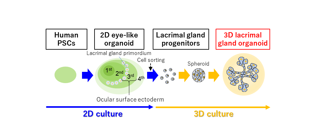
Fig.1 Summary of the study
Ocular surface ectoderm, which is thought to be the common primordium of the lacrimal gland and cornea, is induced in zone-3 of eye-like organoids derived from human iPS cells. After further differentiation culture, we induced and isolated lacrimal gland progenitor cells by cell sorting and successfully generated 3D lacrimal gland organoids by 3D culture in Matrigel.
(credit: Ryuhei Hayashi et al.)
Fig.2 Induction of lacrimal gland progenitor cells and 3D lacrimal gland organoids
(A) Lacrimal gland-like cell clusters induced in zone-3 (lacrimal gland/corneal primordial region) of human iPS cell-derived eye-like organoids were isolated and cultivated in Matrigel, resulting in the formation of 3D lacrimal gland-like organoids with budding and branching. (B) Lacrimal gland organoids were generated by the 3D culture of lacrimal gland progenitor cells isolated by cell sorting. (C) 3D lacrimal gland organoids expressed lacrimal gland markers (PAX6, AQP5, SOX9).
(credit: Hayashi R. et al. Generation of 3D lacrimal gland organoids from human pluripotent stem cells. Nature 2022. Permission to use the content must be obtained at "Rights and Permissions" button that can be found at the bottom of the article available online.)
Fig.3 Transplantation of lacrimal gland organoids into animals
(A) Five lacrimal gland progenitor cell spheroids were cultured in the same well for the preparation of transplantable lacrimal gland organoids (x5). (B) Immunostaining of the transplanted tissues (4 weeks) indicated maturation of the lacrimal gland including ductal tube formation and apical localization of AQP5, which were not observed in vitro. (C) ELISA demonstrated that transplanted lacrimal gland organoids (LGO x1, x5) produce human lactoferrin.
(credit: Hayashi R. et al. Generation of 3D lacrimal gland organoids from human pluripotent stem cells. Nature 2022. Permission to use the content must be obtained at "Rights and Permissions" button that can be found at the bottom of the article available online.)
The article, “Generation of 3D lacrimal gland organoids from human pluripotent stem cells,” was published in Nature at DOI: https://doi.org/10.1038/s41586-022-04613-4.
