About this research

NAGAO Hirofumi
- Graduate School of Medicine
- Endowed Chair Associate Professor (Lecturer)
Elucidated the mechanisms of T-cadherin–mediated regulation of intracellular signaling and its organ-protective effects
- Discovered that T-cadherin suppresses excessive ERK signaling in metabolic organs and cells, thereby exerting organ-protective effects in the heart and skeletal muscle.
- The regulatory mechanisms of T-cadherin signaling in cells and tissues of metabolic organs has not been clarified until now, however, the research group has elucidated it through experiments using cultured cells and mouse models.
- It is expected that a deeper understanding of the onset mechanisms of various metabolic diseases in T‑cadherin–expressing cells and organs will lead to the development of new therapeutic strategies.
Outlines
A research group at the Graduate School of Medicine, The University of Osaka, including Endowed Chair Assistant Professor Hirofumi Nagao (Metabolism and Atherosclerosis), Professor Iichiro Shimomura (Department of Metabolic Medicine), and Endowed Chair Associate Professor Hitoshi Nishizawa (Metabolism and Atherosclerosis), discovered that T‑cadherin, a binding partner of the adipocyte-specific secreted protein adiponectin, regulates intracellular signaling and exerts protective effects on organs such as the heart and skeletal muscle.
The research group has previously reported that adiponectin, an adipocyte-specific secreted factor, binds to the GPI-anchored protein T‑cadherin, enhances exosome production, and exerts a variety of protective effects on multiple organs. While T‑cadherin has been known to regulate intracellular signaling in cancer cells and neurons, its role in cells and tissues of metabolic organs has remained unclear.
In this study, the research group clarified, by using both cultured cells and mouse models, that T‑cadherin suppresses ERK signaling, accompanied by quantitative changes in membrane proteins, including a reduction in IGF‑1 receptor protein levels. Furthermore, in mice genetically engineered to completely lack T‑cadherin in all cells, isoproterenol administration or high-salt loading led to enhanced cardiac hypertrophy, accompanied by excessive ERK activation and an increase in heart weight. It was also revealed that, in mice genetically engineered to lack T‑cadherin specifically in skeletal muscle cells, the physiological muscle atrophy normally induced by fasting was less likely to occur.
These results suggest that T‑cadherin, a principal binding partner of adiponectin, in addition to its reported role in promoting exosome production, regulates intracellular signaling independently of adiponectin and contributes to the maintenance of cellular and organ homeostasis. The findings are expected to deepen the understanding of the onset mechanisms of metabolic diseases in T‑cadherin–expressing organs, including the heart and skeletal muscle, and to lead to the development of new therapeutic strategies.
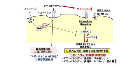
Fig. 1 Regulation of intracellular signaling and organ-protective effects by T‑cadherin
Credit: NAGAO Hirofumi
Research Background
Adiponectin, an adipocyte-specific secreted factor, is known to exert protective effects on multiple organs including the vasculature, skeletal muscle, heart, and kidneys through its anti-atherogenic effect, insulin-sensitizing, and anti-inflammatory actions.
The research group has previously revealed that adiponectin binds to T‑cadherin, a GPI‑anchored protein, leads to increased exosome production and the exertion of diverse protective effects on multiple organs. The research group has also reported that adiponectin enhances T‑cadherin protein expression over the long term.
While it has been known that reduced T‑cadherin expression in cancer cells is linked to tumor progression and poor prognosis, and T‑cadherin suppress neurite outgrowth in neurons, highlighting its role as a regulator of intracellular signaling, its function in cells of metabolic organs has remained unclear.
Research Contents
In this study, the research group clarified through proteomic analyses that T‑cadherin negatively regulates ERK signaling by modulating the abundance of membrane proteins, including the IGF‑1 receptor. This effect was observed not only in cultured cells (F2 endothelial cells and C2C12 myocytes) but also in vivo in mice.
In mice lacking T‑cadherin, cardiac hypertrophy induced by isoproterenol administration or high-salt loading was markedly enhanced, accompanied by excessive ERK activation and an increase in heart weight. Furthermore, in mice with skeletal muscle–specific deletion of T‑cadherin, it was revealed that the physiological muscle atrophy normally induced by fasting was less likely to occur.
Social Impacts
It has been reported that SNPs in T-cadherin, a major binding partner of adiponectin, are associated with metabolic diseases such as metabolic syndrome, obesity, diabetes, and hypertension. In this study, the research group demonstrated that T‑cadherin plays an important role in maintaining cellular and organ homeostasis by regulating intracellular signaling.
Adiponectin itself is not directly involved in this signaling regulatory mechanism; rather, it may support this mechanism upstream by enhancing the protein levels of T‑cadherin. These findings are expected to deepen the understanding of the onset mechanisms of metabolic diseases in T‑cadherin–expressing organs, including the heart and skeletal muscle, and to lead to the development of new therapeutic strategies.
Notes
The article, “T-cadherin, a Major Adiponectin Binding Partner, Suppresses ERK Signaling in Metabolic Tissues,” was published in American scientific journal of Proceedings of the National Academy of Sciences of the United States of America (online) at DOI: https://doi.org/10.1073/pnas.2530597123.