About this research

NAGANO Seiichi
- United Graduate School of Child Development
- Specially Appointed Professor (full-time)
Unraveling the cause of ALS!
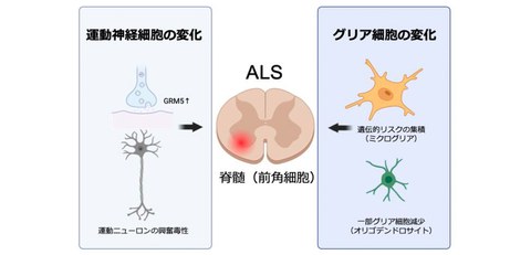
Discovered excessive excitation and dysfunction of cell–cell communication (CCC) in spinal motor neurons
- For the first time in the world, simultaneous analysis of intercellular interactions in ALS patients' brains and spinal cords revealed gene expression changes indicating a state of excessive excitation in spinal motor neurons.
- Furthermore, it was found that the number of cells called oligodendrocytes, which support the neurons, was decreasing, which demonstrated the molecular mechanism that had previously been hypothesized: disrupted cooperation between neurons and other cells influences the onset of ALS.
- It is expected that new treatments for ALS will be developed, such as drugs that suppress excessive motor neuron excitation and therapies that regulate the function of microglia.
Outlines
The research group, including Guest Professor Eriko Takeuchi of the University of Osaka Graduate School of Medicine, Yoshiaki Yasumizu (doctoral student at the time of the research, currently Associate Research Scientist at Yale University), Guest Professor Hideki Mochizuki (Director of the National Hospital Organization Osaka Toneyama Medical Center), and Specially Appointed Professor Shigeo Murayama (full-time) and Specially Appointed Professor Seiichi Nagano (full-time) of the United Graduate School of Child Development at the University of Osaka has revealed for the first time in the world that the gene called GRM5 is overactive in spinal motor neurons of ALS patients, using cutting-edge genetic analysis technology applied to lesion sites from patients with this intractable disease that causes paralysis of motor neurons in the brain and spinal cord. This suggests the excessive activity of the excitatory neurotransmitter glutamate.
Additionally, it was discovered that a portion of the cells called oligodendrocytes, which support neural signaling and nutrient supply, had decreased, disrupting the communication between neurons. Furthermore, it has been discovered that many of the genetic traits that predispose individuals to ALS are concentrated in the brain's immune cells called microglia. These findings revealed that ALS not only causes abnormalities in the motor neurons themselves but is also a disease of the intercellular network in which neurons and surrounding cells influence each other.
Until now, it had been speculated that excessive neuronal excitation caused by glutamate and abnormalities in intercellular networks were involved in the onset of ALS, but the detailed molecular mechanisms had not been elucidated.
In this study, the research group used a single-nucleus multiome analysis to simultaneously observe the brain and spinal cord to clarify which genes are active and the state of each individual cell, and then, elucidated these molecular mechanisms. This is expected to advance the development of new treatments for ALS.
Fig. 1 Changes in the state of each cell in the spinal cord of ALS patients
Credit: Seiichi Nagano
Research Background
ALS, which causes muscle weakness throughout the body due to paralysis of the motor neurons in the brain and spinal cord, is difficult to treat, and there is currently no reliable way to halt the progression of the disease.
Although previous research has shown that in many ALS patients, TAR 2 DNA-binding protein 43 (TDP-43) is lost from the neuronal nuclei and forms abnormal aggregates in the nerves, it was not well understood why motor neurons are so prone to damage and how cells other than neurons are involved.
Conventional genetic analysis could only provide average information for the entire tissue, making it impossible to accurately examine differences between individual cells. Therefore, the research team analyzed all cells in the brain and spinal cord at a single-nucleus level to investigate in detail which cells were changing and how in ALS.
Research Contents
1. Created a map of the brain and spinal cord that covers all cells
The research group analyzed approximately 130,000 cells from the brains and spinal cords of ALS patients and those without motor neuron disease, and classified various cell types, such as neurons and glial cells (cells that support the neurons). This data is valuable on a global scale.
2. Discovered abnormal excitation in motor neurons
In the motor neurons of the spinal cord of ALS patients, a gene called GRM5 was abnormally active. This gene is a blueprint for making glutamate receptors, which strengthen the signals that excite neurons. In other words, excitotoxicity, a state in which the motor neurons remain overly excited, occurs and this is thought to be one of the factors of nerve damage.
3. Decrease in cells protecting neurons
In the spinal cord, there was a decrease in some of the oligodendrocytes, which support the function of neurons. These cells create the myelin sheath that surrounds neurons and play important roles such as transmitting information within neuron cells and supplying nutrients. The loss of myelin sheath is thought to make neurons more prone to weakening.
4. Genetic factors are concentrated in immune cells
Genetic changes that increase susceptibility to ALS were frequently observed in microglia, immune cells in the brain. This provides new evidence supporting the influence of the immune response in the progression of ALS.
Social Impact of Research
These findings are expected to lead to the development of drugs that suppress excessive excitation of motor neurons (drugs targeting GRM5) and treatments that regulate the function of microglia. Furthermore, the perspective on this disease will change, and not only neurons but also the cells supporting them may become important targets for treatment.
The analytical data obtained in this study will be made available to researchers around the world, significantly advancing research into ALS and related neurological disorders.
Notes
The article, “Single-Nucleus Multiome Shows Motor Neuron Glutamate Overactivation in Amyotrophic Lateral Sclerosis,” was published in British scientific journal Brain at DOI: https://doi.org/10.1093/brain/awaf426.