
Visualizing the whole-brain rhythmicity in three dimensions
- Neural activities throughout the mouse brain were analyzed by using the tissue clearing technology CUBIC, and it was clarified that approximately 80% of the brain region has a circadian rhythmicity.
- The research group has expanded the analysis, which was previously limited to certain brain regions, to the whole brain level under highly reproducible conditions, and highly accurate time-series analysis was conducted to comprehensively visualize rhythms throughout the brain.
- The results of this study provide a new foundation for understanding brain function from time-series perspective and is expected to contribute to the development of time-dependent research and medical applications, such as those into sleep, memory, and drug efficacy.
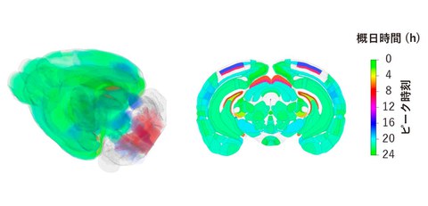
Three-dimensional whole-brain rhythmicity map of a mouse
Credit: Katsunari Yamashita
Outlines
A research group including Professor Hiroki Ueda of the Graduate School of Medicine, The University of Tokyo (also serves as a Distinguished Invited Professor at Kurume University), Special Research Student Katsunari Yamashita (a doctoral student at the Graduate School of Medicine, The University of Osaka), Project Researcher Fukuaki Kinoshita of the University of Tokyo, and Associate Professor Rikuhiro Yamada of the Institute of Life Sciences, Kurume University, has clarified that neural activity in various regions of the mouse brain indicate circadian rhythmicity.
In this research, by using the tissue clearing technology CUBIC to observe neural activity throughout the mouse brain tissue collected over time, the researchers demonstrated, for the first time in the world, that approximately 80% of the brain region exhibits a circadian rhythmicity. While previous research has focused on limited brain regions, this study comprehensively evaluated the entire brain using high-precision time-series analysis and made the results available online as a whole-brain database. The results of this study provide a new foundation for understanding brain function from time-series perspective and is expected to contribute to the development of time-dependent research of brain function and medical applications, such as those into sleep, memory, and drug efficacy.
Research Contents
Our brain manages various functions, including sleep, arousal, memory, and emotions, in accordance with the time flow of a day. Previous research has clarified that a small region of the brain called the suprachiasmatic nucleus is the center of controlling the body's internal clock, and it has been reported that some of such brain regions exhibit the rhythm of neural activities that follow a cycle of a day. Electrical recording and measurement of gene expression have been used to observe neural activity. However, the area which could be observed was spatially limited, making it impossible to capture how whole-brain activity changes throughout the day. Furthermore, spontaneous neural activity in the brain is easily affected by slight environmental differences and external stimuli, making it difficult to accurately measure changes throughout the day under highly reproducible conditions.
The research group analyzed 144 mouse brains collected sequentially over two days under constant darkness using CUBIC, a tissue clearing technology that makes organs transparent and allows for three-dimensional observation of internal structures, and c-Fos immunostaining, which visualizes neural activity (Fig. 1). By combining strict set conditions that minimize environmental impacts with highly accurate image and rhythm analysis methods, the researchers have achieved time-series evaluation of spontaneous neural activity, which was difficult until now.
Fig. 1. Overview of the method of experiment
Mouse brains were collected sequentially over two days under constant darkness, and changes in whole-brain neural activity were analyzed using tissue clearing and three-dimensional immunostaining.
The analysis demonstrated that not only the suprachiasmatic nucleus, but also approximately 80% (508 regions) of the 642 regions that make up the brain exhibit circadian rhythmicity (Fig. 2). Mice are nocturnal, and many regions indicate peaks during the latter half of the night (active period) according to the body's internal clock. On the other hand, regions related to vision and sleep showed peaks during the day (inactive period) according to the body’s internal clock. In the hippocampus, which is responsible for memory, the CA1 region and the dentate gyrus are active at different times in turns, suggesting that even within the regions control a range of functions, there is a division of rhythms according to their roles. Furthermore, voxel-level rhythm analysis clarified that different activity rhythms exist even within a single region (Fig. 3).
Fig. 2 Changes in neural activity in brain regions showing circadian rhythmicity
The suprachiasmatic nucleus showed peak activity during the daytime according to the body’s internal clock, same as previous reports. In the hippocampus, which is responsible for memory, activity peaks in the CA1 and dentate gyrus appeared at opposite times. Also, activity peaks in multiple areas related to various physiological functions, such as arousal and vision, were concentrated at specific time ranges. Across the brain, 508 of 628 regions (approximately 79%) indicated circadian rhythmicity.
Fig. 3 Whole-brain three-dimensional rhythm analysis in voxel units (suprachiasmatic nucleus)
The research team analyzed whole-brain activity rhythms in voxel units. Even within a single region, peak activity in different time zones was observed.
Furthermore, the research group showed that it is possible to estimate the time of day (brain time) from the whole-brain activity pattern (Fig. 4). These results suggest that whole-brain activity patterns have a possibility to be used as a new index for measuring brain condition.
Fig. 4 Estimating the time of day from whole-brain activity patterns
The whole-brain activity patterns, based on the neural activity of each brain region (the ratio of the number of c-Fos-positive cells to the whole brain), showed unique patterns at different times of day. Taking advantage of this characteristic, the research group was able to estimate the time on the body's internal clock (brain time) with high accuracy from a single brain sample.
The data obtained has been made available online as a 3D whole-brain rhythm atlas, and anyone can freely access it. This database will be a foundation for functional research into various brain regions and is expected to be applied to research into understanding brain function from a time axis and analyzing the effects of drugs depending on the time of day.
Notes
The article, “A whole-brain single-cell atlas of circadian neural activity in mice,” was published in Science at DOI: http://www.science.org/doi/10.1126/science.aea3381.