
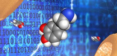
Rapid detection of neurotransmitters using single molecular measurements and machine learning
A group of researchers from The Institute of Scientific and Industrial Research and the Graduate School of Frontier Biosciences of Osaka University has developed a method to detect neurotransmitters at the single molecule level by applying single molecule measurements to analysis using machine learning (ML) on the millisecond time scale with a time resolution of 10 millisecond (ms).
Studies of neurotransmitters, which are associated with neurological disorders, including depression and Parkinson's disease, are essential for the understanding of nerve action. To understand these disorders, detailed measurements of neurotransmitter interplay with good temporal resolution is required.
As measures to identify single molecules, there is a technique based on nanoelectrodes for electrically detecting and distinguishing single molecules with good temporal resolution. To discriminate a single molecule, tunneling current flowing through a single molecule (or conductance) is measured using nanogap electrodes with a gap of 1 nm or less. This provides information on the electronic state of the molecule, resulting in the detection of the individual electrical conductivity. Gaps in the sub-10 nm range can be produced by techniques, such as electromigration (EM)-induced breaking of thin metal wires.
This group developed a method to identify a single molecule using a tunneling current flowing between the modified electrodes through a single analyte molecule. When sample molecules pass between nanogap electrodes, electron transfer occurs as a quantum mechanical tunnel effect.
In single molecule measurements, the conductance of molecules between the gaps is directly measured, so multiple types of neurotransmitters can be detected simultaneously in a single nanogap. However, conventional methods can hardly identify the target molecules selectively and accurately because the current signals include various noise signals from other neurotransmitters, contaminants, and the migration of the gold electrodes and single molecule conductance is easily affected by them.
To solve this noise problem, the researchers developed an analysis method based on machine learning (ML) from the waveform of the tunneling current flowing between nanoelectrodes via a single-molecule. They removed these noise signals using a positive and unlabeled data (PU) classifier, a ML algorithm for distinguishing between two classes when only positively labelled and unlabeled data are given.
Using this method, they enhanced the classification performance. They measured neurotransmitters in different parts of the mouse brain and identified dopamine, serotonin, and norepinephrine neurotransmitters.
The method developed by this group will allow one to observe neurotransmitters traveling across the synaptic cleft at the site detected by single molecule measurements and to perform highly selective measurements of neurotransmitters with high temporal resolutions. This will lead to detailed measurements of neural activity, which will also enable direct measurements of neurotransmitters between synapses.
These developments will lead to a deep understanding of disorders associated with neurotransmitters and the discovery of new medications to treat such disorders. This group’s new analysis method will significantly increase the scope of application of single molecular measurements and will be applied to measurements of various molecules.
Figure 1
The article, “Time-resolved neurotransmitter detection in mouse brain tissue using an artificial intelligence-nanogap”, was published in Scientific Reports at DOI: https://www.nature.com/articles/s41598-020-68236-3.