
Nearly double resolution of conventional microscopes achieved
Will improve the quality and functionality of products in a wide range of industrial fields
A team of researchers led by FUJITA Katsumasa (Professor, Graduate School of Engineering), KAWATA Satoshi (Professor, Graduate School of Engineering), and HASHIMOTO Hitoshi (Professor, Graduate School of Pharmaceutical Sciences) at Osaka University succeeded in nearly doubling the conventional resolution of Raman scattering microscopy, which is used in a wide variety of fields in science and industry ranging from drug discovery to device development.
Raman scattering is a very subtle effect, so it was thought difficult to increase the resolution of Raman scattering microscopy. However, this group succeeded in measuring at high spatial resolution by shedding dashed light on samples, achieving the highest resolution of microscopy using propagating light.
This group’s research results have made it possible to observe spatial distribution of drugs and device materials in further detail, which is expected to be helpful in enhancing the quality and functionality of products.
Unlike conventional super-resolving microscopy, which requires dying of samples with pigment, the technique developed by this group does not require this dying of samples. This technique will become an important milestone in the development of microscopy.
Abstract
In the last couple of decades, the spatial resolution in optical microscopy has increased to unprecedented levels by exploiting the fluorescence properties of the probe. At about the same time, Raman imaging techniques have emerged as a way to image inherent chemical information in a sample without using fluorescent probes, however, in many applications, the achievable resolution is limited to about half the wavelength of excitation light. Here we report the use of structured illumination to increase the spatial resolution of label-free spontaneous Raman microscopy, generating highly detailed spatial contrast from the ensemble of molecular information in the sample. Using structured line illumination in slit-scanning Raman microscopy, we demonstrate a marked improvement in spatial resolution and show the applicability to a range of samples, including both biological and inorganic chemical component mapping. This technique is expected to contribute towards greater understanding of chemical component distributions in organic and inorganic materials.

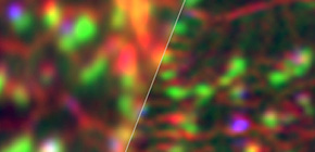
Raman images of CVD graphene. Structured line illumination (left) provides higher spatial resolution compared to that by conventional line illumination.
Fig.1 Line illumination used in conventional slit-scanning Raman microscopy (left) and structured line illumination used for the developed Raman microscope (right).
Fig.2 Raman images of CVD graphene. Red, blue and green indicate Raman scattering from D, G, and 2D band of the graphene. Structured line illumination visualized distribution of single/double layer graphene, graphite and defect with higher spatial resolution. The plot at the right bottom shows the line profiles of Raman signal assigned to D band measured between the arrows in the images.
Fig.3 Raman images of a mouse brain slice. Red and green indicate Raman scattering from molecular vibrations of peptide (rich in proteins) and CH 2 (rich in lipids), respectively. Distribution of lipids is more clearly seen by structured line illumination. The plot at the right bottom right shows the line profiles of Raman signal assigned to CH 2 stretching vibration measured between the arrows in the images.
To learn more about this research, please view the full research report entitled “ Structured line illumination Raman microscopy ” at this page of the Nature Physics website.
Related links