
High-speed, high-resolution spinning disk confocal microscopy for intravital imaging
Riken and Osaka University have developed a method enabling high-speed and high-resolution spinning disk confocal microscopy of thick specimens such as living tissues and animals.
This research achievement was realized in cooperation with the following: Yuko MIMORI-KIYOSUE , Unit Leader, and SHIMOZAWA Togo , Researcher, Researcher Optical Image Analysis Unit, Center for Developmental Biology (CDB), Riken; FUJITA Katsumasa , Associate Professor, Graduate School of Engineering, Osaka University; YAMAGATA Kazuo , Specially Appointed Associate Professor, Research Institute for Microbial Diseases, Osaka University; KONDO Takeshi , Special Postdoctoral Researcher, Laboratory for Morphogenetic Signaling, CDB, Riken; SHITAMUKAI Atsunori and KONNO Daijiro , both Research Specialists, Laboratory for Cell Asymmetry CDB, Riken; TAKAYAMA Jun , Postdoctoral Researcher, Laboratory for Developmental Dynamics, Quantitative Biology Center, Riken; and WATANABE Tomonobu , Team Leader of Laboratory for Comprehensive Bioimaging. Joint research groups at Yokogawa Electric Corporation and Coherent Japan, Inc . also participated in this research.
Imaging technology to enable visualizing the behavior of living molecules and cells is essential for life science research. Although technology to view behavior of fast-moving molecules inside cells has made significant progress, the ability to view molecurels in relatively deep regions in living organisms has remained ellusive. This joint research group targeted the development of a new imaging technique through the improvement of existing devices and methods.
This group succeeded in improved spinning disk confocal microscopy by increasing interpinhole distance (thereby significantly reducing image cross-talk) and by employing two-photon excitation that made possible selective-plane illumination. Making use of these, they found that in regions 100µm deep in specimens, the contrast ratio increased 30 times or more over that of conventional confocal microscopy.
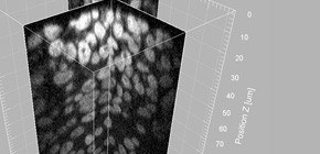
Furthermore, this group succeeded, for example, in live imaging of an elongated microtubule (25nm in diameter) necessary for maintaining and deforming the shape of cells in regions 25µm deep in the drosophila embryo, demonstrating high-speed and high-resolution imaging of deep regions in living organisms.
This device will likely contribute to advances in a wide range of life sciences including basic research, application research, and medical treatment.
Abstract
A recent key requirement in life sciences is the observation of biological processes in their natural in vivo context. However, imaging techniques that allow fast imaging with higher resolution in 3D thick specimens are still limited. Spinning disk confocal microscopy using a Yokogawa Confocal Scanner Unit, which offers high-speed multipoint confocal live imaging, has been found to have wide utility among cell biologists. A conventional Confocal Scanner Unit configuration, however, is not optimized for thick specimens, for which the background noise attributed to “pinhole cross-talk,” which is unintended pinhole transmission of out-of-focus light, limits overall performance in focal discrimination and reduces confocal capability. Here, we improve spinning disk confocal microscopy by eliminating pinhole cross-talk. First, the amount of pinhole cross-talk is reduced by increasing the interpinhole distance. Second, the generation of out-of-focus light is prevented by two-photon excitation that achieves selective-plane illumination. We evaluate the effect of these modifications and test the applicability to the live imaging of green fluorescent protein-expressing model animals. As demonstrated by visualizing the fine details of the 3D cell shape and submicron-size cytoskeletal structures inside animals, these strategies dramatically improve higher-resolution intravital imaging.

Figure 1

Figure 2

Figure 3

Figure 4
To learn more about this research, please read the full research report entitled " Improving spinning disk confocal microscopy by preventing pinhole cross-talk for intravital imaging " at this page of the Proceedings of the National Academy of Sciences of the United States of America (PNAS) website.
Related links: