
Pulling the Genome Apart: Chromosome Segregation During Mitosis Explained
Researchers reveal the importance of a pathway in controlling the segregation of chromosomes when cells divide, which could be useful for treating a range of diseases associated with abnormal cell division
When a cell divides—a process known as mitosis—its chromosomes need to be separated and evenly distributed into the newly created daughter cells. Although this is known to be extremely complicated and to feature a range of cellular components, many of its details remain unclear, which has hampered efforts to develop treatments for when mitosis goes awry.
A new study reported in the journal Nature Cell Biology has provided deeper insight into this process by revealing the details of how protein complexes congregate at the centers of chromosomes, at sites known as centromeres. At these sites, the protein complexes act as anchors through which cellular structural organizers can redistribute chromosomes within the cell.
These protein complexes on centromeres are known as kinetochores, to which long, thin, cylindrical structures called microtubules attach. In cell division, microtubules can physically manipulate the chromosomes, pulling half of each chromosome for inclusion into one daughter cell, and half into the other.
In this new study,the Osaka University-led team focused on the different components that form and bind to kinetochores. One such group is the CCAN proteins, which are present at the centromere throughout the cell cycle and act as a binding site for other microtubule-associating proteins only when cell division occurs. This work shows that a subset of the CCAN proteins, forming the CENP-T pathway, dominates in ensuring successful cell division by binding to a protein complex called the Ndc80 complex, enabling microtubules to attach to chromosomes.
“We chiefly investigated whether the CENP-T pathway or the roughly similar CENP-C pathway is essential for mitotic progression by selectively deleting parts of these proteins that bind to the Ndc80 complex,” corresponding author Tatsuo Fukagawa says. “CENP-T mutants with an absence of domains for binding to Ndc80, but not similar CENP-C mutants, revealed a failure of chromosomes to segregate, preventing cells from dividing and ultimately leading to cell death.”
To confirm their results and reveal more about CENP-T’s essential role in promoting cell division via the Ndc80 complex, the team also constructed chimeric constructs consisting partly of CENP-T and partly of CENP-C. This validated the finding that CENP-T and the molecules that bind to it are vital for mitosis. They also revealed that phosphorylation plays a vital role in regulating binding between the Ndc80 complex and CENP-T, and obtained additional direct evidence for their findings by measuring the pulling force exerted by microtubules in the mitotic spindle.
“Our work overturns an earlier consensus by showing that it is CENP-T, not CENP-C, that acts via the Ndc80 complex for successful cell division by ensuring accurate and timely chromosome segregation,” lead author Masatoshi Hara says. “The findings could lead to therapeutic options for treating diseases involving dysfunction in the kinetochores and mitotic progression, including cancer.”
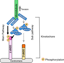
Fig. 1: A model of how spindle microtubules bind to the kinetochore.
To establish a functional kinetochore, the microtubule-binding Ndc80 complex (Ndc80C) must be recruited into the kinetochore. While a previous model suggested that CENP-C is a main recruiter for Ndc80C, this study demonstrates that CENP-T is actually the main player in Ndc80C recruitment into the kinetochore.
(credit: Osaka University)
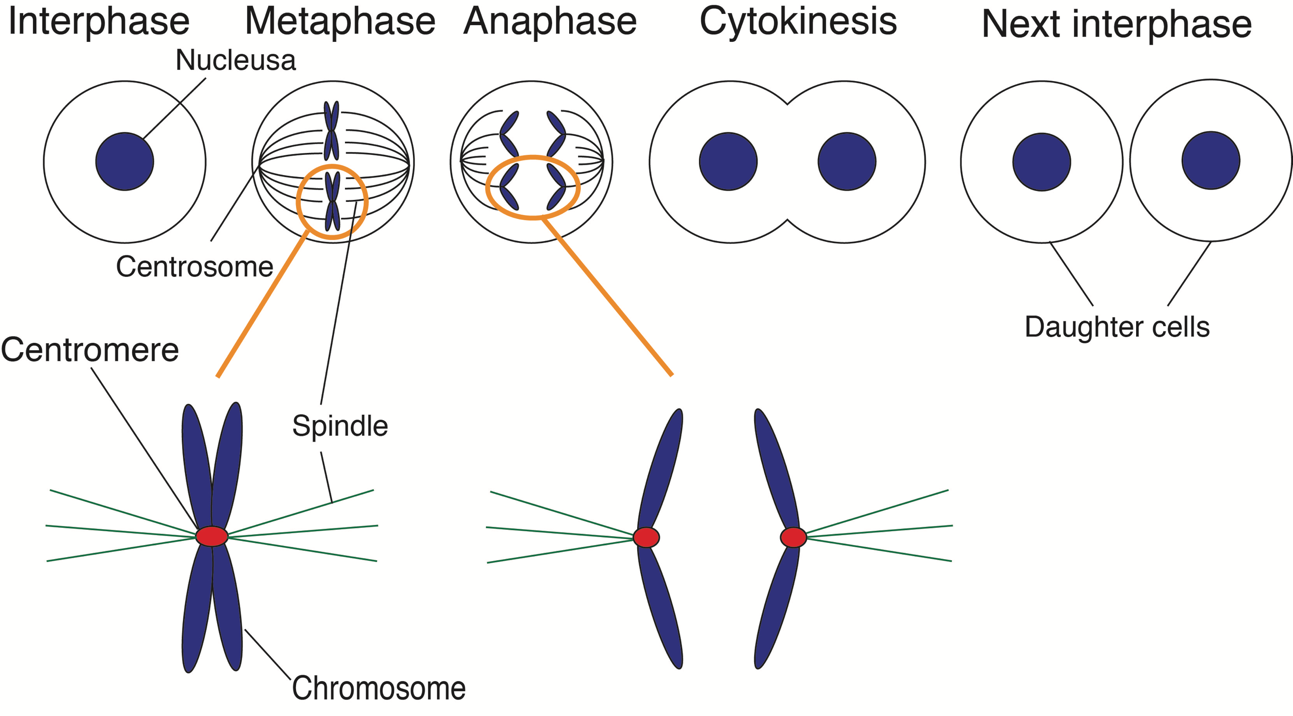
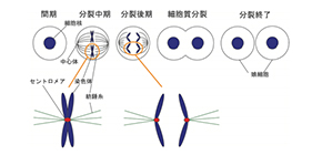
Fig. 2: Schematic presentation of mitotic progression.
During mitosis, spindle microtubules capture the kinetochore formed in the centromere. After mitosis, chromosomes are divided into daughter cells.
(credit: Osaka University)
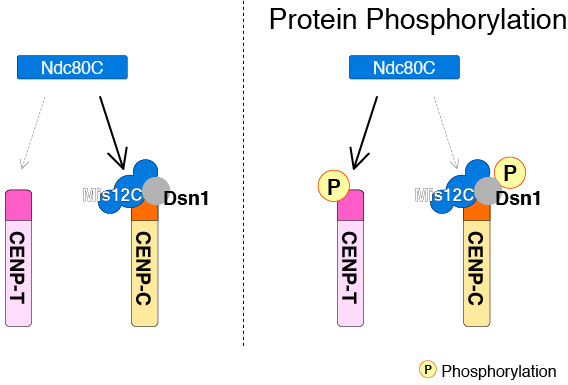
Fig. 3: A model of how Ndc80 preferentially binds to CENP-T rather than CENP-C.
In the absence of protein phosphorylation, Ndc80C preferentially binds to CENP-C-containing complex. Once protein phosphorylation occurs, Ndc80C preferentially binds to CENP-T.
(credit: Osaka University)
The article “Multiple phosphorylations control recruitment of the KMN network onto kinetochores” is published in Nature Cell Biology at DOI: https://doi.org/10.1038/s41556-018-0230-0 .
Related links