
Micro-RNA separation by nanopillars and nanoslits technique
Will lead to application to single cell analysis and nanopore sequencing
A group of researchers led by Professor BABA Yoshinobu and Associate Professor KAJI Noritada at the Graduate School of Engineering, Nagoya University, together with Specially Appointed Professor KAWAI Tomoji at The Institute of Scientific and Industrial Research, Osaka University, Professor YANAGIDA Takeshi at Kyushu University, and Professor TOKESHI Manabu at Hokkaido University, succeeded in separation of only microRNA (miRNA), which is thought to be a promising biomarker, from nucleic acids at the very high speed of 20 milliseconds (ms) by using nanopillars and nanoslits technique.
MiRNAs are approximately 22-nucleotide-long, non-coding RNA molecules that are not translated into a protein. In recent years, it is suggested that miRNAs are highly correlated with development of cancer and are drawing attention as easy-to-acquire, nearly non-invasive potential biomarkers for the diagnosis and prognosis of cancer.
MiRNA extraction was performed by conventional solid phase extraction using silica beads; however, it required samples of tens of micron liters or bigger and manual handling. These problems kept miRNAs from being applied to a pretreatment for semiconductor nanopore sequencing that enables reading base sequences directly from a single DNA or RNA molecule.
By using a microfabrication technique used for semiconductor manufacturing, this group fabricated nanobiodevices in which nanopillar structures that have nanometer-scale pillar structures are combined with nanoslit structures. With this hybrid structure of nanopillars and nanoslits, this group improved the speed of separation of nucleic acids. The process of separation and extraction of miRNAs, which conventionally took tens of seconds, was achieved within 100 ms.
This group also succeeded in extracting let-7, a biomarker for cancer, within only 20 milliseconds. This nanobiodevice enables extraction of miRNAs from a sample of a couple of picoliter (= 10-12 liter). Thus, by extracting desired miRNAs from a single cell and using them in combination with a device such as nanopore-based DNA sequencer that is capable of reading base sequences of nucleic acids at a single molecular level, this technique will realize full-scale single-cell analysis.
Abstract
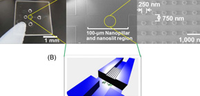
A millisecond micro-RNA separation of a mixture of total RNA and genomic DNA, extracted from cultured HeLa cells, was successfully achieved using a hybrid structure of nanopillars and nanoslits contained inside a microchannel. The nanopillars, 250-nm in diameter and 100-nm in height, were fabricated with a 750-nm space inside the nanoslits, which were 100-nm in height and 25-μm in width; the nanopillars were then applied as a new sieve matrix. This ultra-fast technique for the separation of miRNA can be an effective pretreatment for semiconductor nanopore DNA sequencing, which has an optimum reading speed of 1 base/ms to obtain effective signal-to-noise ratio and discriminate each base by ion or tunneling current during the passage of nucleic acids.
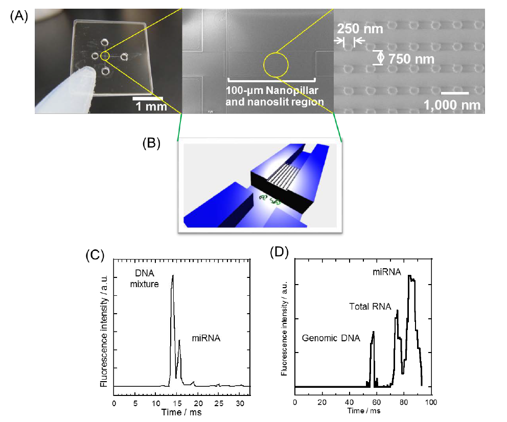
Figure 1
To learn more about this research, please view the full research report entitled " A millisecond micro-RNA separation technique by a hybrid structure of nanopillars and nanoslits " at this page of the Scientific Reports website.
Related link