

Mechanism of spontaneous recovery from CNS damage in MS discovered
As part of JST's basic research programs, under the leadership of MURAMATSU Rieko , Assistant Professor and YAMASHITA Toshihide , Professor at the Graduate School of Medicine, Osaka University, a group of scientists have succeeded in experiments using mice in clarifying a central nervous system (CNS) regenerative mechanism pertinent to therapy for multiple sclerosis (MS) and in the speeding up of remission from neural damage.
In MS, CNS damage by inflammation causes severe symptoms such as numbness and blurred vision and such symptoms undergo both remission and recurrence. It has been thought that recovery from CNS damage was difficult because of weak regenerative ability of the CNS. Nonetheless, it has been noted that partial spontaneous recovery takes place in damaged nerve axons. Likewise, it has been thought such neural regeneration can lead to remission in MS, but the mechanism for such has been unknown.
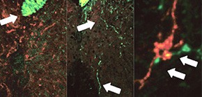
This group discovered that the formation of new blood vessels plays a part in axonal rewiring, leading to functional recovery by examining tissues of mice with experimental autoimmune encephalomyelitis (EAE), similar to MS, and clarified that prostacyclin, a protein from blood vessel endothelial, increased the regeneration ability of axons.
This group has also succeeded in halving the time for recovery from EAE mice by strengthening the function of prostacyclin. This research has clarified for the first time in the world that angiogenesis functions in neuronal rewiring and in increasing the regenerative power of the CNS. Their findings will lead to the possible development of new drugs targeting functional recovery from CNS damage and multiple sclerosis.
This research was conducted in cooperation with MOCHIZUKI Hideki , Professor, Graduate School of Medicine, Osaka University, and FUJIMURA Harutoshi , Chief, Clinical Research Department, Toneyama National Hospital.
Abstract
" Angiogenesis is a prominent feature of central nervous system (CNS) disease and has roles in both the continued promotion of inflammation and the subsequent repair processes. Here we report that prostacyclin (or prostaglandin I2 (PGI2)) derived from new vessels promotes axonal remodeling of injured neuronal networks after CNS inflammation. In a localized model of experimental autoimmune encephalomyelitis (EAE), new vessels formed around the inflammatory lesion, followed by sprouting of adjacent corticospinal tract (CST) fibers. These sprouting fibers formed a compensatory motor circuit, leading to recovery of motor function. Capillary endothelial cell–derived prostacyclin bound to its receptor, the type I prostaglandin receptor (IP receptor), on CST neurons, promoting sprouting of CST fibers and contributing to the repair process. Inhibition of prostacyclin receptor signaling impaired motor recovery, whereas the IP receptor agonist iloprost promoted axonal remodeling and motor recovery after the induction of EAE. These findings reveal an important function of angiogenesis in neuronal rewiring and suggest that prostacyclin is a promising molecule for enhancing functional recovery from CNS disease. "
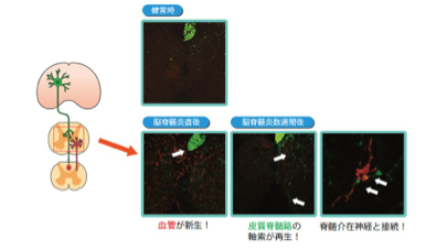
Figure 1
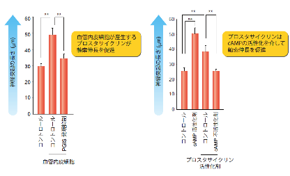
Figure 2
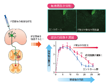
Figure 3
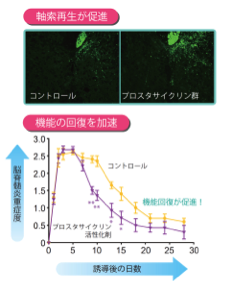
Figure 4
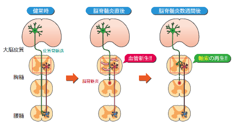
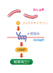
Figure 5
To read the full research report entitled " Angiogenesis induced by CNS inflammation promotes neuronal remodeling through vessel-derived prostacyclin , " please go to this page at the Nature Medicine website.
Related links :