
\Uncovering how hidden retinal blood vessels are protected/ Discovery of the retinal vascular stem cell reservoir in the optic nerve
Toward a new therapy that promotes vascular repair in retinal disorders
- The researchers revealed that a reservoir of stem/progenitor cells that supplies vascular endothelial cells responsible for forming the retinal vasculature—an essential tissue for vision, exists in the optic nerve.
- Retinal blood vessels are essential for vision, yet it has long been unknown where new endothelial cells come from in adults or which cells repair the vessels when they are damaged.
- The findings of this study are expected to lead to new therapeutic treatments that can physiologically rebuild the capillary networks damaged in ischemic retinal disorders, including diabetic retinopathy, retinopathy of prematurity, and retinal vein occlusion.
Outlines
A research group including Professor Koji Nishida and Endowed Chair Associate Professor Susumu Sakimoto at the Graduate School of Medicine, the University of Osaka has elucidated that stem/progenitor cells supplying retinal vascular endothelial cells are abundantly present in the optic nerve. While some cell-surface markers of tissue-resident vascular endothelial progenitor/stem cells have been reported, and these cells were believed to be mainly located in large vessels, their exact location and how they supply new cells had remained unknown.
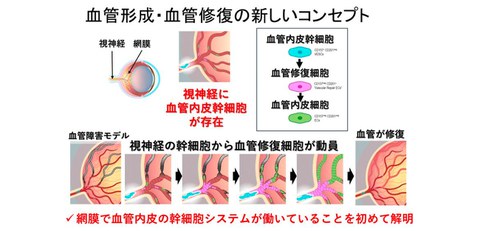
In this study, the research group used single-cell RNA sequencing (scRNA-seq) analysis in mice to identify vascular endothelial stem/progenitor cells that accumulate in the optic nerve. Through lineage tracing and a damaged blood vessel model (oxygen-induced retinopathy), it was demonstrated that these cells are supplied from the optic nerve to the retina and are mobilized for repair upon damage. In addition, these findings showed that the optic nerve contains a reservoir of stem cells that supports the maintenance and repair of the retinal blood vessel (Fig. 1).
These findings are expected to the development of new therapeutic targets and regenerative strategies that restore the physiological capillary networks lost in ischemic retinal disorders, including diabetic retinopathy, retinopathy of prematurity, and retinal vein occlusion.
Fig. 1 Summary of research findings
The study identified that stem cells of retinal blood vessels reside abundantly in the deeper optic nerve and identified a new model in which cells are mobilized from stem cells for repair upon damage to rebuild blood vessels
Credit: Koji Nishida
Research Background
The retina is a neural tissue that supports vision and requires a large amount of oxygen and nutrients. Therefore, prolonged interruption of retinal blood flow results in impaired visual function. Diseases such as diabetic retinopathy, retinopathy of prematurity, and retinal vein occlusion cause damage to the retinal blood vessels, leading to reduced vision and blindness.
Until now, only a few cell-surface markers of tissue-resident vascular endothelial progenitor/stem cells had been reported, and these cells were believed to be located mainly in large blood vessels. However, the detailed mechanisms underlying where retinal vascular endothelial cells are routinely supplied in adults and which cells are responsible for vascular repair following ischemic injury had remained unknown.
Research Contents
By using mouse models, the research group focused on the cell-surface markers CD157/Bst1 and CD201/PROCR, which are known as indicators of vascular endothelial stem/progenitor cells, and analyzed vascular endothelial cells in the retina and optic nerve.
While CD157‑positive cells represented only approximately 1–2% in the retina, the optic nerve contained a markedly higher proportion, with over 20% of the stem/progenitor cells indicating CD157 and CD201. These results indicate that optic nerve–derived vascular endothelial cells display robust colony-forming capabilities and exhibit characteristics consistent with stem/progenitor cells (Fig. 2).
Furthermore, scRNA‑seq analysis (Fig. 3), together with lineage tracing and a damaged blood vessel model (Fig. 4) clarified that under ordinally conditions, progenitor cells in the optic nerve replenish retinal vascular endothelial cells, and that following damage, a specific progenitor population (CD201‑positive cells) becomes activated and contributes to vascular repair. These findings were supported by the finding that deletion of the transcription factor ATF3 in progenitor cells resulted in an expanded avascular area during the repair phase, clarified that progenitor cells are essential for the restoration of the physiological capillary network.
Fig. 2 Vascular endothelial stem cells exist in the optic nerve
(a) Top: Schematic illustration of the optic nerve and retina
(b) FACS analysis of CD157 and CD201, markers of endothelial stem cells, indicated over a 20‑fold increase in CD157‑positive and CD201‑positive cells relative to the retina in the optic nerve
(c) In the vascular endothelial colony‑forming assay, colonies were formed from CD157‑positive/CD201‑positive cells
Credit: Koji Nishida
Fig. 3 Single‑cell RNA sequencing analysis of vascular endothelial cells in the retina and optic nerve
(a) Experimental schema
(b) Clusters from CD45‑negative/CD31‑positive cells
(c) Clusters expressing Bst1 (CD157), Procr (CD201), and Lrg1-positive were observed
(d) These stem cell clusters were absent in samples containing only the retina and emerged only when the optic nerve was included
Credit: Koji Nishida
Fig. 4 Lineage tracing using Procr (CD201)‑CreERT2-tdTomato mice
(a) Under ordinally conditions, tdTomato‑positive cells were observed only in the optic nerve at two days after tamoxifen administration, but by day seven they appeared in the central retinal veins, and by day 14 they extended to the peripheral veins
(b) Analysis in the oxygen‑induced retinopathy (OIR) damaged blood vessel model. At postnatal day 13, no tdTomato‑positive cells were observed in the retina; however, by postnatal days 17 and 28, as the vasculature underwent repair, most of them were repaired by tdTomato‑positive cells
Credit: Koji Nishida
Social Impacts
This study indicates that the optic nerve contains a reservoir that maintains and repairs the retinal blood vessel, and it highlights the potential for developing new therapies aimed at regenerating retinal blood vessels. In particular, in diseases such as diabetic retinopathy, where pathological neovascularization following ischemic changes poses a major clinical problem, the ability to restore the physiological capillary network could help reduce the risk of severe complications, including hemorrhage and retinal detachment.
Notes
The article, “Endothelial stem cells of the retinal vasculature reside in the optic nerve,” was published in American journal of Nature Communications (online) at DOI: https://doi.org/10.1080/23294515.2025.2474928.