About this research

MATSUNO Kenji
- Center for Overseas Operations, Institute for International Initiatives
- Specially Appointed Professor

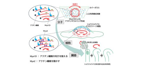
Mechanism determining right- and left-handed chirality
Unraveling chirality across molecules, cells, and the body through the actin chiral ring (ACR)
- The researchers revealed the molecular mechanism underlying the clockwise intracellular flow of actin filaments, a key component of the cellular cytoskeleton.
- Although chirality has long been recognized at the molecular, cellular, and organ levels in living organisms, how molecular chirality gives rise to chirality at higher levels of biological organization remains unclear.
- Using fluorescence live-cell imaging, the researchers discovered that actin filaments flow clockwise within cells under the action of the motor protein Myosin 1D (Myo1D). They also found that Myo1D bundles actin filaments into a clockwise-rotating ring structure, termed the “actin chiral ring (ACR),” even on a glass substrate.
- The result provides important insights into the fundamental mechanisms governing chirality in living organisms. The findings are expected to advance research on organogenesis and organoid development, while also contributing to an elucidation of tissue robustness and the mechanisms underlying cancer invasion.
Outlines
A research group including Specially Appointed Researcher Asuka Yamaguchi (at the time of the study), Visiting Associate Professor Takeshi Sasamura (at the time of the study), Professor Kenji Matsuno (at the time of the study; currently Professor Emeritus at the University of Osaka) of the Department of Biological Sciences, Graduate School of Science, The University of Osaka, together with researchers from Chiba University, the University of Toyama, Kyoto University, and RIKEN, has, for the first time in the world, elucidated the mechanism by which cells acquire right- and left-handed chirality.
Although the human body appears externally symmetrical, its internal organs display a distinct chirality, with the heart, for example, positioned on the left side of the body.
Understanding how organs are positioned and develop with the correct orientation during embryonic development has been a central question in developmental biology. This property, known as cellular chirality, plays an important role in establishing left–right asymmetry during organogenesis. However, the mechanisms that determine whether a cell becomes right- or left-handed have remained unclear.
In this study, the research team observed living Drosophila cells and performed a detailed analysis of the dynamics of actin filaments, which form the cellular cytoskeleton. As a result, the team discovered that when Myosin 1D (Myo1D), an actin-based motor protein known to be involved in chirality in animals, is active, actin filaments flow through the cell in a clockwise direction (Fig. 1, cellular level).
The research team reconstituted this intracellular phenomenon in vitro using only essential molecular components, including Myo1D and actin. Then it was discovered that Myo1D induces the formation of an actin chiral ring (ACR), in which numerous actin filaments assemble and rotate in a clockwise direction (Fig. 1, molecular level). Because the ACR is comparable in size to a cell, a similar mechanism may operate within cells to generate a clockwise flow of actin filaments. This finding indicated a fundamental mechanism by which Myo1D generates the clockwise flow of actin filaments within cells.
These findings provide a foundation for future studies aimed at elucidating the fundamental principles governing chirality in animals, including humans, and determining how cellular chirality contributes to tissue destabilization and cancer invasion.
Fig. 1 Overview of the mechanism underlying cell chirality formation
Credit: Kenji Matsuno
Research Background
Living organisms, including humans, exhibit clear chirality in the shape and positioning of internal organs such as the heart, stomach, and liver. How such chirality of the body and internal organs is established is one of the fundamental questions in developmental biology.
Recent studies have revealed that the properties underlying such chirality are also present at the level of individual cells. Cells that constitute organs and tissues possess intrinsic left-right biases in their morphology, migration, and intracellular behavior. This is known as “cell chirality,” because it exhibits chirality, a property in which an object and its mirror image cannot be superimposed, much like the relationship between the right and left hands. Cell chirality has attracted increasing attention in recent years, as it has been implicated not only in the formation of organs exhibiting chirality but also in the maintenance of tissue robustness and, when disrupted, in the invasion of cancer cells. However, the mechanisms that determine whether cells exhibit right- or left-handed chirality have remained unclear.
Research Contents
Using Drosophila macrophages, which are well suited for microscopic observation, the research team visualized the movement of actin filaments in living cells and discovered that the flow of actin filaments is directed clockwise. To study the mechanism underlying this clockwise flow, the researchers focused on Myo1D, a motor protein known to play a crucial role in establishing right- and left-handed chirality in a variety of organisms, including humans, zebrafish, and Drosophila. These results revealed that Myo1D plays a key role in orienting actin filament flow toward the clockwise direction (Fig. 1, cellular level). Furthermore, the researchers found that this rotational movement also requires another myosin motor protein, Myosin2 (Myo2).
The researchers then sought to reproduce this cellular phenomenon in a simplified experimental system. To this end, they immobilized Myo1D on a glass substrate and added actin filaments at concentrations comparable to those found inside cells, allowing them to observe their behavior in an artificial reconstitution experiment. The result revealed that Myo1D induces the bundling of actin filaments into ring-shaped structures that undergo clockwise rotational motion (Fig. 1, molecular level). These self-organized rotating actin assemblies are known as an actin chiral ring (ACR).
Individual actin filaments have an intrinsic polarity, defined by a plus end and a minus end. Furthermore, Myo1D is a motor protein that transports actin filaments exclusively in the direction of their minus ends. These observations suggest that the unidirectional rotation of ACRs arises from the coordinated alignment of the constituent actin filaments. Thus, Myo1D functions as an organizer that aligns the polarity of multiple actin filaments and drives their collective motion in a single direction (Fig. 1, molecular level). This is thought to be the mechanism underlying the clockwise intracellular flow of actin filaments. However, why the direction reverses and becomes counterclockwise in the absence of Myo1D remains unclear and will be an important subject for future investigation.
Based on these findings, the research team proposed a model for the establishment of cellular chirality in which Myo1D aligns the polarity of actin filaments, while Myo2 drives their movement, thereby generating the clockwise intracellular flow of actin filaments. Because Myo1D plays a key role in determining right- and left-handed chirality in the digestive tract and reproductive organs of Drosophila, the model proposed in this study is thought to represent a fundamental mechanism underlying the establishment of the chirality in organs and tissues (Fig. 1, organ level).
Thus, this study revealed how molecular-level right- and left-handed chirality, exemplified by the Myo1D-driven ACR, gives rise to the overall right- or left-handedness of a cell.
Social Impact
These findings make a significant contribution to the understanding of the mechanism underlying the establishment of cell chirality, a process essential for the development of the body plan in living organisms. Building on these findings, future applications are expected not only in studies of organogenesis, regeneration, and organoid development, but also in pathological research, including investigations into how cellular chirality contributes to tissue destabilization and cancer invasion.
Notes
The article, “Myosin1D directs circumferential F-actin flow to define cell chirality,” was published in American scientific journal of Journal of Cell Biology at DOI: https://doi.org/10.1083/jcb.202509125.