About this research

SAITO Takuro
- Graduate School of Medicine
- Specially Appointed Associate Professor (Full Time)

Unraveling the mechanisms of immunotherapy in gastrointestinal cancers
Anti-PD-1 antibodies target progenitor-exhausted T cells in lymph nodes!
- The research group demonstrated that treatment with immune checkpoint inhibitors (ICIs) induces the proliferation of lymph node–resident progenitor-exhausted T cells, leading to activate anti-tumor immunity in patients with gastrointestinal cancers.
- It had long been thought that reactivation of exhausted T cells within the tumor microenvironment was essential for the efficacy of ICI therapy. However, by analyzing, at the single-cell level, T cells directly bound by anti-PD-1 therapeutic antibodies after ICI treatment, the research group found that immune cells in lymph nodes, as well as those within tumors, play a critical role in the therapeutic response.
- Because the study suggests the effectiveness of ICI therapy when administered in the preoperative setting, while lymph nodes are still preserved, it is expected to develop new treatment strategies for gastrointestinal cancers. These may include the development of immunotherapies that specifically target progenitor-exhausted T cells within lymph nodes.
Outlines
A research group of the University of Osaka including Yohei Nose, Visiting Academic Staff at the Graduate School of Medicine, Takuro Saito, Specially Appointed Associate Professor (Full Time) at the Department of Clinical Research in Tumor Immunology and Department of Gastroenterological Surgery, and Yuichiro Doki, Specially Appointed Professor at the Department of Gastroenterological Surgery, has demonstrated for the first time in the world that progenitor-exhausted T cells residing in regional lymph nodes play a crucial role in treatment with immune checkpoint inhibitors (ICIs) for gastrointestinal cancers.
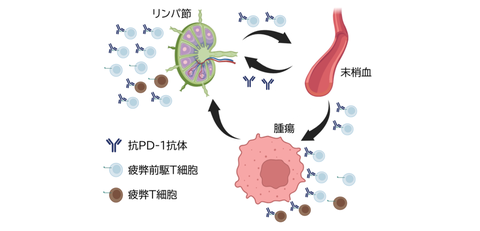
Until now, anti-PD-1 antibody therapy, a type of ICI treatment, has been thought to exert its antitumor effects by reactivating exhausted PD-1-positive T cells present within the tumor microenvironment. However, studies using experimental animal models have suggested that these ICI therapies may exert their effects on immune cells within the lymph nodes (Fig. 1).
In this study, the research group performed a single-cell analysis of T cells directly bound by therapeutic anti-PD-1 antibodies reside in tumors, lymph nodes, and peripheral blood from patients with gastrointestinal cancers who underwent surgery after anti-PD-1 antibody treatment. As a result, the study revealed that progenitor-exhausted T cells in the lymph nodes proliferate and undergo clone expansion in response to ICI therapy, subsequently differentiating into exhausted T cells within the tumor, and exert antitumor effects. These findings demonstrate that lymph nodes, in addition to the tumor microenvironment, are critical sites for the therapeutic efficacy of anti-PD-1 antibodies.
These findings suggest that administering ICI therapy in the preoperative setting, when lymph nodes are still preserved, may be particularly effective for the treatment of gastrointestinal cancers. The study is expected to contribute to the development of future therapeutic strategies for gastrointestinal cancer.
Fig. 1 Schematic overview of this study
Anti-PD-1 antibodies increase the number of progenitor-exhausted T cells not only within tumors but also in lymph nodes and promote their recruitment to tumors.
Credit: Takuro Saito
Research Background
In recent years, anti-PD-1 antibody therapy, a type of immune checkpoint inhibitor (ICI) treatment, has become a mainstay of therapy for patients with unresectable advanced gastric and esophageal cancers. When tumor cells express PD-L1, it binds to PD-1 expressed on T cells, thereby suppressing T-cell activity. Conventionally, anti-PD-1 antibodies have been thought to exert their therapeutic effects by blocking the binding between PD-L1 and PD-1 within the tumor microenvironment, thereby reactivating suppressed T cells and enabling them to attack tumor cells (Fig. 1).
On the other hand, recent studies using experimental animal models have reported that ICIs act on T cells not only within the tumor microenvironment but also in lymph nodes. However, it has remained unclear how ICI therapy influences immune cells in tissues outside the tumor microenvironment, such as lymph nodes and peripheral blood. This study aims to elucidate the effects of ICI therapy on immune cells throughout the body by analyzing samples obtained from patients who underwent surgery after receiving ICI treatment.
Research Contents
Anti-PD-1 antibodies are thought to act on T cells that express PD-1. First, the researchers performed flow cytometric analysis of T-cell subsets in tumor tissues, metastatic lymph nodes, and non-metastatic lymph nodes (pLNs, proximal lymph nodes; dLNs, distal lymph nodes) from 55 ICI-naïve patients with gastric cancer. Specifically, the proportions of progenitor-exhausted T cells (Tpex; TCF1⁺PD-1⁺CD8⁺ T cells), exhausted T cells (Tex; TCF1⁻PD-1⁺CD8⁺ T cells), and PD-1⁻CD8⁺ T cells were evaluated.
The analysis revealed that Tpex cells were enriched in non-metastatic pLNs, whereas Tex cells were predominantly found within tumors (Fig. 2). Furthermore, patients with a high Tpex/Tex ratio in non-metastatic lymph nodes indicated a favorable prognosis (Fig. 3), suggesting that the presence of Tpex cells in non-metastatic lymph nodes may play a critical role in clinical outcomes.
Next, the research group investigated how these T-cell populations change following ICI therapy. Because anti-PD-1 antibodies exert their effects by binding to PD-1-expressing T cells, therapeutic antibodies are thought to be directly bound to PD-1-positive T cells. In this study, the research group took advantage of the fact that anti-PD-1 antibodies belong to the IgG4 subclass and established a method to identify T cells bound by therapeutic antibodies using an anti-IgG4 antibody (Fig. 4). Using this method, the researchers analyzed samples from patients who underwent surgery after ICI therapy and evaluated the proliferative capacity of each T-cell subset, as assessed by Ki-67 expression, in tumors, metastatic lymph nodes, and non-metastatic lymph nodes. The analysis revealed that, following ICI therapy, the proliferative capacity of Tpex cells was markedly enhanced, particularly in non-metastatic lymph nodes (Fig. 5).
Fig. 2 Proportions of Tpex, Tex, and PD-1⁻CD8⁺ in different tissues from patients with gastric cancer
Credit: Takuro Saito
Fig. 3 Tpex/Tex ratio in non-metastatic lymph nodes and prognosis
Kaplan–Meier curves of recurrence-free survival (RFS) according to the Tpex/Tex ratio in non-metastatic lymph nodes of patients with gastric cancer
Credit: Takuro Saito
Fig. 4 Detection of PD-1⁺ cells
Credit: Takuro Saito
Fig. 5 Proliferative capacity of cell population before and after ICI treatment
The frequency of Ki-67-expressing cells among Tpex, Tex, and PD-1⁻CD8⁺ T-cell populations in different tissues from patients with and without ICI treatment
Credit: Takuro Saito
Furthermore, by integrating single-cell RNA-seq, CITE-seq, and TCR repertoire analyses, the research group characterized therapeutic antibody-bound T cells at the single-cell level in each tissue after ICI treatment (Fig. 6). T cells from tissue samples obtained after ICI treatment were subjected to clustering analysis, which identified a cell population corresponding to lymph node Tpex cells (Cluster 5). This cell population was shown to undergo clone expansion while remaining directly bound by the therapeutic anti-PD-1 antibody. Furthermore, analysis of TCR similarity revealed that ICI-bound Tpex cells in lymph nodes shared a high proportion of TCR clones with intratumoral Tex cells (Clusters 4 and 6). These findings suggest that lymph node Tpex cells proliferate in response to ICI treatment and subsequently differentiate into Tex cells within the tumor.
Fig. 6. Integrated single-cell RNA-seq, CITE-seq, and TCR repertoire analysis of post-ICI treatment samples
(a) Clustering analysis of T cells isolated from tumor, liver metastases, lymph nodes, and peripheral blood of two gastric cancer patients who underwent surgery after ICI treatment (b) Distribution of clusters across different tissues (c) Degree of clone expansion across clusters (d) Network analysis of TCR similarity among each cluster
Credit: Takuro Saito
Social Impacts
These findings provide mechanistic insights into how ICI therapy exerts its antitumor effects and identify lymph nodes as a critical target organ for its therapeutic activity. They also support the efficacy of administering ICI therapy in the preoperative setting, when lymph nodes are still preserved, in the treatment of gastrointestinal cancers. They also prompt a reconsideration of the optimal timing for ICI administration.
Notes
The article, “PD-1 antibody–bound progenitor-exhausted CD8⁺ T cells in lymph nodes boost PD-1–blockade anti-tumor immunity in gastrointestinal cancer,” was published in British journal of Nature Communications at DOI: 10.1038/s41467-026-70751⁻2.
