Lasers light up neutron generation for radiography
Researchers from Osaka University report a laser-driven neutron source for acquiring non-destructive radiography images
Getting snapshots of systems and processes at precise time points is important to research and development in many fields, including biology, materials science, and engineering. Firing a neutron beam at a material is one way of gaining information; however, this often requires nuclear reactors and specialist facilities. Now, researchers from Osaka University have reported a laser-driven method of simultaneously generating neutrons and X-rays. Their findings are published in Applied Physics Express.
Looking at a system without having to destroy it is very useful when investigating different structures. One way this can be done is to aim light, ionizing radiation, or particles at the material of interest and see how they interact with the target.
Neutrons—particularly low-energy ones—are excellent particles for this because they are likely to interact with different nuclei, including hydrogen. However, generating neutrons can require specialist facilities that are not easily accessible.
Recently, systems using lasers to generate neutrons have been gaining popularity because they are compact, can generate short bursts of neutrons, and can produce X-rays at the same time.
The Osaka researchers have developed a laser-driven neutron source that is small—the size of a fingertip—and can generate a lot of fast neutrons in very short bursts. The neutrons are then slowed down by a moderator to make them optimal for imaging.
“We were able to generate a high neutron density—higher than is found in some stars—which means we could acquire the information needed very rapidly,” explains study corresponding author Associate Professor Akifumi Yogo. “X-rays were also produced at the same time, so the system can offer two complimentary techniques in one.”
The neutrons were generated by switching a laser on and off. This control over the neutron source makes the system safer than previous alternatives.
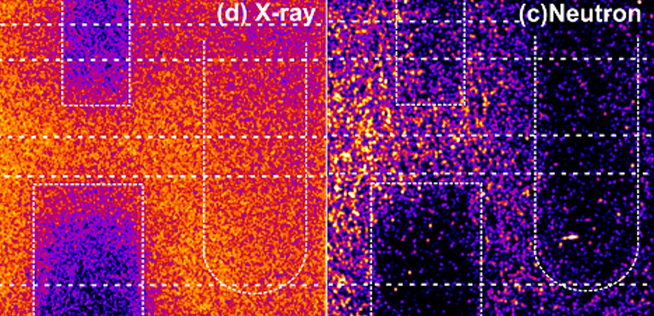
The researchers used their technique to show that boron carbide, which could not be imaged using X-rays, was detected using neutrons. In addition, they examined hazardous substances in a typical battery in a non-destructive way, and were able to detect the presence of cadmium using neutrons.
“The rapid neutron burst we were able to achieve with our system will provide imaging information for very rapid processes,” says Associate Professor Yogo. “For example, we believe events such as fuel injection in engines and bubble collapse in fast jets could be observed, which would provide valuable information for research in many industries.”i

Fig.1 The results of “snap shot” radiography using laser-induced neutrons and X-rays simultaneously. (Left) Photograph of the samples: Nickel metal hydride battery (Ni-MH), nickel cadmium battery (Ni-Cd), and boron carbide powder (B2C). (Center) X-ray radiography, where B2C is transparent to X-rays. (Right) Neutron radiography. The Ni-Cd can be distinguished from the Ni-MH based on the darkness of the shadow. In addition, low transmittance was observed for B2C. These results highlight the advantage of neutrons, which can identify materials that are transparent to X-rays.
(credit: © 2021 A. Yogo et al., Applied Physics Express)
The article, “Single shot radiography by a bright source of laser-driven thermal neutrons and x-rays,” was published in Applied Physics Express at DOI: https://doi.org/10.35848/1882-0786/ac2212.
