
Optical imaging of muons will be useful for precise measurement of muon beam and elementary particle research
A group of researchers from Nagoya University, OsakaUniversity, and the Institute of Materials Structure Science (IMSS) of the High Energy Accelerator Research Organization (KEK), has measured muon beam distribution using optical imaging.
Muons, elementary particles, can be produced in mass by an accelerator and high-energy cosmic muons, which penetrate matter, are used for the radiography of a volcano dome, pyramid, or nuclear reactor.
Elemental analysis and material properties research using high intensity pulse muon beams generated at accelerators can achieve accurate results during a short period of time.
Optical imaging is a promising approach for quality assessment (QA) of high energy X-rays from linear accelerators (LINAC) and mainly measures Cherenkov-light from electrons or secondary electrons produced in water. Cherenkov light is generated when a charged particle such as an electron and a positron move through matter like water faster than the speed of light.
High intensity muon beams can be used at some facilities such as the Japan Proton Accelerator Research Complex (J-PARC). Recently, the luminescence of water at a lower energy than the Cherenkov-light threshold for particle ions was found and has been used for dose and range estimations. Luminescence of water was found in X-ray, alpha particles, and beta particles and optical imaging for dose and range estimation was tried for these radiations, but it has not been tried for muons.
To conduct elemental analysis and material properties research, obtaining information about how muon beams hit on a target, distribution of muon beams, and how muon beam energy spreads is important. If imaging of muon beams and positrons emitted from muons when they decay is available, it will become possible to get information from images.
Assuming that it would be possible to image high-intensity muon beams using a cooled charge-coupled device (CCD) camera at J-PARC, the researchers focused on Cherenkov-light, conducting experiments.
When positively charged muon beams pass through water, only positrons emit Cherenkov light, which is produced by the decay of muons. This group obtained images of Cherenkov light for 300 s during muon beam irradiation. The elliptical light distributions in the images were produced by Cherenkov light of positrons in the water from the decay of positive muons.
The researchers irradiated positive muons with different momenta to water or plastic scintillator block, and imaged using a CCD camera during irradiation. The depths of the peaks of the distributions for measured and simulated images were similar; both peaks were located deeper for muons with higher momenta.
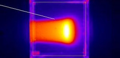
For the plastic scintillator block, which emits sufficient fluorescent light upon exposure to radiation, the researchers conducted imaging for 10 s and obtained light distributions having Bragg peaks, which occur just before the particle comes to a complete stop.
From these images, this group obtained information about beam energy and estimate beam ranges and widths. Optical imaging will be applied to a wide range of fields, including precision measurements of muon beams and studies of elementary particles.
Figure 1
Figure 2
Figure 3
The article, “Optical imaging of muons,” was published in Scientific Reports at DOI: https://www.nature.com/articles/s41598-020-76652-8.