Learning how cancer cells coordinate and collaborate to multiply and metastasize
Researchers from Osaka University and the National Institute of Information and Communications Technology show that cancer cells cultured on Matrigel migrate to form a network structure as they do in vivo, and describe the forces responsible
Cancer cells are known to migrate and collaborate to form networks that function as conduits providing access to nutrients and blood vessels. Now, researchers in Japan have generated similar large-scale structures from cancer cells in the laboratory and thus gained a better understanding of the underlying forces and their interactions.
Proliferating cells often cooperate in order to form self-beneficial large-scale structures; these include bacterial biofilms, protective epithelial monolayers or even more complex configurations such as endothelial capillaries. Malignant cells, in a process called vasculogenic mimicry, form structures that facilitate access to nutrients for tumor growth and to blood vessels for metastasis. The biochemical and biophysical mechanisms are not well understood, as this purposive behavior was difficult to reproduce experimentally until now.
In a study published in the Biophysical Journal in March 2020, researchers from Osaka University in collaboration with Advanced ICT Research Institute, the National Institute of Information and Communications Technology (NICT), have demonstrated migration and large-scale structure formation by cancer cells grown on Matrigel substrate, and have developed simple simulated models that reproduce their observations.
The research team first cultured HeLa cells, a strain of epithelial-like cervical cancer cells, on Matrigel, a gelatinous protein mixture resembling the extracellular environment of many tissues, and showed that the cells migrate aggressively and form large-scale structures. This was previously difficult to achieve in vitro as HeLa cells are relatively non-motile on glass. Using time-lapse imaging they analyzed the cell migration patterns and quantified the large-scale structures with a two-point correlation function.
“We observed that HeLa cells first exhibited increased motility on Matrigel, which later decreased after they integrated into a spatially distinct structure,” explains Dr. Tokuko Haraguchi, senior researcher at NICT. “We also noted that HeLa cells in close proximity formed bridges between cell aggregates, and that structures were formed in a cell-density dependent manner.”
To explain these results, the researchers developed a simulated model in which cells migrate and interact using two distinct forces: remote forces that act at a distance through substrate deformation, and contact forces between cells in physical proximity. By selectively enabling these forces, they modelled the three types of structures formed—islands, network-like structures and continents—according to cell density.
Tadashi Nakano, lead author, explains the potential implications of their findings. “Cancer cells may rely on vasculogenic mimicry for survival and proliferation,” he says. “A complete understanding of this process can, by manipulation of the relevant cell density and force parameters, inhibit these network-like structures. This may have great potential for combating cancer.”

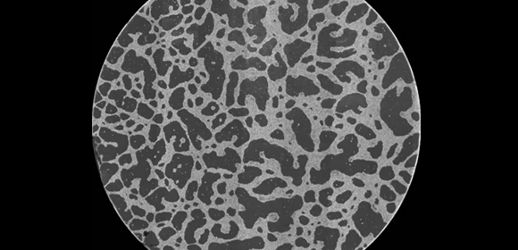
Fig.1 A large-scale network structure formed in vitro by cancer cells. The diameter of the image is 14 mm.

Fig.2 Collective migration of cancer cells and formation of cell aggregates. The leftmost image shows the initial distribution of cancer cells. Approximately 700 cells are shown in the image. The remaining three images together show the 5-hour trajectories of 71 randomly selected cells. A number of cells have moved toward the upper-right position in the images and formed an aggregate. The area of each image is 1 mm x 1 mm.

Fig.3 Cellular bridge formation to connect two cell aggregates. The cell in the green circle initially moved to the bottom-left position (0-1 hour), and then extended its body toward the cell in the blue circle (2.5-5 hours). As a result, the cell ‘bridged’ two cell aggregates, one at the bottom left and the other at the top right. The area of each image is 0.5 mm x 0.5 mm.
The article, “Roles of remote and contact forces in epithelial cell structure formation” was published in the Biophysical Journal at DOI: https://doi.org/10.1016/j.bpj.2020.01.037 .
Related links
