
A New Technique for the Observation of Membrane Lipid Distribution
Successful observation performed by attaching a small tag to the target lipid
A group of researchers led by Michio Murata (Professor at the Graduate School of Science, Osaka University), and Mikiko Sodeoka (Director, Synthetic Organic Chemistry Laboratory, RIKEN) observed the distribution of membrane lipid in artificial monolayer by the introduction of a small tag, alkyne, to the target lipid. The lipid was considered to be of uniform distribution at raft-like ordered domain, but it was heterogeneous. Their developed methodology of alkyne-tag Raman imaging enables them to perform these observations. These results are expected to contribute to a new analysis and an elucidation of cell membrane lipid.
Abstract
Membrane micro-domains called lipid rafts have been known to be involved in a variety of important biological events such as signal transduction, protein sorting and virus infection. Since the raft, extracted as the detergent resistant membrane (DRM) fraction from biomembranes, contains large amounts of sphingolipids represented by sphingomyelin (SM), SM is thought to be a key constituent of the raft. Thus, the distribution of SM in membranes should be essential information to elucidate the mechanism for the raft formation and for cite specific biological events. So far, fluorescent lipid analogs have been employed to visualize the distribution of the target lipids. However, almost all fluorescent SM analogs showed the different disposition from natural SMs probably because a large fluorophore linked to the target lipid causes the perturbation of lipid packing and modulates the membrane properties. In this sense, we newly synthesized Raman-tagged SM (diyne-SM) where a small diyne moiety is attached to the SM polar head and employed ‘hyperspectral’ Raman microscopy developed by Fujita et al. (Department of Applied Physics, Osaka University) to visualize the distribution of diyne-SMs because the diyne moiety provides a relatively strong peak at the Raman silent window.
Consequently, we successfully obtained the Raman image showing the distribution of diyne-SM in an artificial-raft supported monolayer comprising of SM/dioleoylphosphatidylcholine/cholesterol (1:1:1 by mole) ternary mixture. Moreover, the Raman images disclosed the gradual decrease of the concentration of SM from the center of the artificial raft domain to the peripheral areas. The gradual change in the concentration of diyne-SM implies a slightly different view from that of the common raft model, in which the raft and non-raft phases undergo a relatively clear biphasic separation. Our approach can provide both chemical selectivity and quantitative imaging capability, and is useful for functional studies of lipid rafts.
Distributional analysis of Diyne-Sphingomyelin
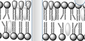
A schematic illustration for a traditional lipid raft model.
In cell membranes, the lipids show the heterogeneous distribution and form specific membrane domains together with certain proteins. Especially, the sphingomyelin-rich domain called lipid raft has been attracting researchers’ interests because it is known to be involved in the important biological event. In the common raft model, the SM-rich raft domain and the SM-poor non-raft matrix undergo a relatively clear biphasic separation.
To learn more about this research, please view the full research report entitled " Sphingomyelin distribution in lipid rafts of artificial monolayer membranes visualized by Raman microscopy " at this page of the Proceedings of the National Academy of Sciences of the United States of America website.
Related Links