Technique developed for the creation of gold nanoparticles at desired intracellular locations
Enables non-invasive measuring of cellular biological phenomena
A group of researchers led by Nicholas Isaac Smith (Associate Professor, Immunology Frontier Research Center [iFReC], Osaka University) have succeeded in fabricating gold nanoparticles (crystals) with laser irradiation of gold ions inside cells.
Using gold nanoparticles in cells produced by this method and Surface Enhanced Raman Scattering (SERS) makes it possible to measure the chemical environment of the gold particles. This technique will be used for non-invasive measuring of biological phenomena in cells in the future.
Various biological reactions and chemical reactions occur inside the cell at the same time. There are several methods to obtain such information. A typical method is to view cells using artificial pigments or fluorescent proteins such as green fluorescent protein (GFP) with a fluorescent microscope.
Prof. Smith has developed a method for obtaining intracellular chemical information through a Raman spectrometric method without the use of pigments or proteins. Unlike the fluorescent microscope, the Raman spectrometric method is expected to be used for measuring not only the appearance of cells, but also chemical information, and the state of intracellular locations.
This group of researchers have succeeded in:
1. creating pure gold nanoparticles by irradiating gold ions inside cells with a 532 nm laser. The diameter of the nanoparticle sizes ranged from 2 to 20 nm and the particles remained in place even after the gold solution had been removed.
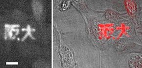
2. precisely controlling gold nanoparticle fabrication. As proof of that, this group succeeded in using a laser focal scan patternf for the characters for 阪大 [Handai, the Japanese nickname for Osaka University], 15 microns in size, fabricated from the gold nanoparticles in the cell.
3. measuring the chemical state of the area surrounding the gold particles by exciting gold nanoparticles fabricated inside cells with 785 nm wavelength irradiation.
This group has provided a new means for gaining intracellular biological information. Their technique can generate gold crystalline structures (nanoparticles) where desired inside the cell through the use of laser irradiation. As SERS enhances Raman signals on the surface of nanoparticles, it becomes possible to obtain precise information about the chemical state of the area surrounding the gold particles. This group's technique will also enable researchers to obtain information about locations of choice in the cell without damaging the cell.
Abstract
Nanoparticle manipulation is of increasing interest, since they can report single molecule-level measurements of the cellular environment. Until now, however, intracellular nanoparticle locations have been essentially uncontrollable. Here we show that by infusing a gold ion solution, focused laser light-induced photoreduction allows in situ fabrication of gold nanoparticles at precise locations. The resulting particles are pure gold nanocrystals, distributed throughout the laser focus at sizes ranging from 2 to 20 nm, and remain in place even after removing the gold solution. We demonstrate the spatial control by scanning a laser beam to write characters in gold inside a cell. Plasmonically enhanced molecular signals could be detected from nanoparticles, allowing their use as nano-chemical probes at targeted locations inside the cell, with intracellular molecular feedback. Such light-based control of the intracellular particle generation reaction also offers avenues for in situ plasmonic device creation in organic targets, and may eventually link optical and electron microscopy.
Figure 1

Figure 2

Figure 3: Phase and spectroscopic imaging of photofabricated characters in gold inside cells

Figure 4
To learn more about this research, please view the full research report entitled " Laser-targeted photofabrication of gold nanoparticles inside cells " at this page of the Nature Communications website.
Related link :
