
Research reveals for the first-time the 3D structure of a membrane protein to form a continuous strand that could be common in the PMP-22/EMP/MP20/Claudin superfamily
Under the leadership of TSUKITA Sachiko , Professor, Graduate School of Frontier Biosciences and Graduate School of Medicine, Osaka University, and FUJIYOSHI Yoshinori , Professor, Cellular and Structural Physiology Institute (CeSPI), Nagoya University, a group of researchers have succeeded in revealing, for the first time, the 3D structure of a membrane protein such as those found in the claudin superfamily of proteins (major cell-adhesion molecules of tight junctions) and in obtaining insight into tight junction adhesion processes.
Living organisms maintain a homeostatic balance by forming a boundary between their bodies and their environment. Epithelial tissue is responsible for forming a protective barrier between the body/cells and the environment by linking its cells in a cell-to-cell adhesion process referred to as tight junction (TJ). Previously, the shape and binding mechanism of claudin and similar proteins has been unknown; however, this group succeeded in revealing the 3D structure of a claudin-like protein, providing strong evidence of a similar structure in TJ strands that mainly comprise claudin proteins.
If the structure of claudin proteins can be further clarified in the near future, this will contribute to the development of therapeutics for inflammatory bowel diseases, such as Crohn disease, in which insufficient TJ adhesion plays a role.
Abstract
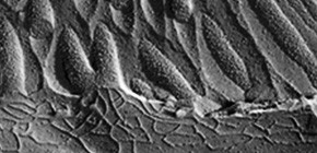
Euglenoid flagellates have striped surface structures comprising pellicles, which allow the cell shape to vary from rigid to flexible during the characteristic movement of the flagellates. In Euglena gracilis, the pellicular strip membranes are covered with paracrystalline arrays of a major integral membrane protein, IP39, a putative four-membrane-spanning protein with the conserved sequence motif of the PMP-22/EMP/MP20/Claudin superfamily. Here we report the three-dimensional structure of Euglena IP39 determined by electron crystallography. Two-dimensional crystals of IP39 appear to form a striated pattern of antiparallel double-rows in which trimeric IP39 units are longitudinally polymerised, resulting in continuously extending zigzag-shaped lines. Structural analysis revealed an asymmetric molecular arrangement in the trimer, and suggested that at least four different interactions between neighbouring protomers are involved. A combination of such multiple interactions would be important for linear strand formation of membrane proteins in a lipid bilayer.

Figure 1

Figure 2
To learn more about this research, please read the full research report entitled "The four-transmembrane protein IP39 of Euglena forms strands by a trimeric unit repeat" at this page of the Nature Communications website.
Related links :