
Modeling the human eye in a dish
Researchers from Osaka University develop a novel human stem cell model to study eye development, biology and disease in a dish
Despite its small size relative to the rest of the body, the eye is one of the most complex organs of the human body and has been difficult to study in a lab. Now, researchers from Osaka University have developed a novel method to model eye development and disease using human induced pluripotent stem cells (hiPSCs). In a new study published in Journal of Biological Chemistry , they showed how tracking the expression of PITX2, a key protein during eye development, in developing hiPSCs enables the isolation of a certain group of cells that play important roles in eye development, biology and disease.
Ever since their discovery over a decade ago, hiPSCs have continued to be used to replicate human biology and disease in a lab without the need for animals. Their streamlined use is accompanied by the possibility of easily genetically altering the cells to study the function of proteins. Although to date several cellular models of multiple organs have been developed using hiPSC, due to its complex and heterogeneous nature, the eye has been more difficult to recreate using these cells.
“Unlike other organs, the eye is more difficult to recreate in the lab due to the presence of heterogeneous cells in the eye,” says corresponding author of the study, Ryuhei Hayashi. “The goal of our study was to develop a novel human cellular eye model using hiPSCs that will help improve our understanding of how these different cell types develop to form the eye.”
To achieve their goal, the researchers established a reporter cell line by modifying hiPSCs using genome editing technology, such that the cells express the fluorescent protein eGFP whenever they express the protein PITX2. PITX2 is a transcriptional factor protein that plays a key role during embryonic development of several organs, including the eyes. In the eye, PITX2 is specifically expressed in what is called periocular mesenchyme (POM), a collection of cells that give rise to the cornea, as well as muscle cells and connective tissue within the eye. As a result, by using the genetically modified cells, the researchers were able to fluorescently label POM cells.
“We wanted to know whether our new cellular model was able to recreate elements of normal eye development and isolated POM cells for characterization,” says lead author of the study Toru Okubo.
The researchers first showed that the modified hiPSCs remained pluripotent after genome editing, so they still maintained the properties of pluripotent stem cells in the same way as unchanged hiPSCs. They then induced the development of POM cells from hiPSCs and showed that they formed so-called self-formed ectodermal autonomous multi-zones (SEAM), which are two-dimensional tissues consisting of different eye cells that form during normal eye development (first reported by Hayashi’s group in 2016). Previously, there were no methods to isolate POM cells, but this new generation of gene-edited iPSCs enables the team to isolate POM cells selectively from the SEAM. By isolating the fluorescent POM cells from other, non-fluorescent cells, the researchers were then able to show that POM cells maintained known molecular markers during further cell culture, validating the recreation of eye development using their hiPSC reporter cell line.
“These are striking results that show how human stem cells can be used to study development and disease processes,” says Hayashi. “Our model could offer a new opportunity to understand how different aspects of eye development happen.”
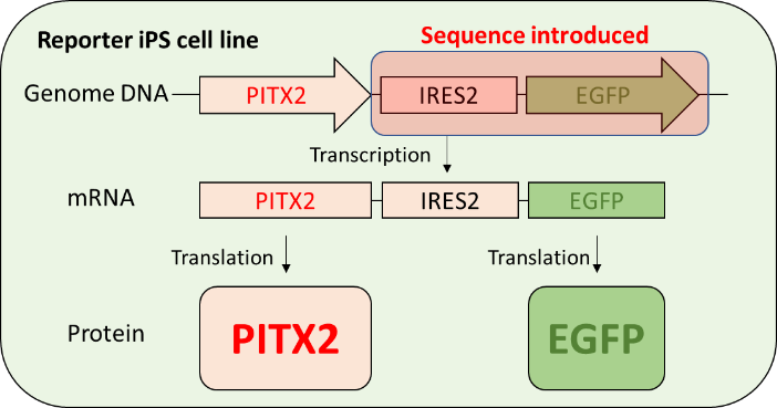
Fig.1 Design of reporter iPS cell line.
IRES2 sequence and EGFP gene were introduced at the downstream of PITX2 gene in the genome of human iPS cells using genome editing technology. EGFP is expressed according to the expression of PITX2 gene expression.
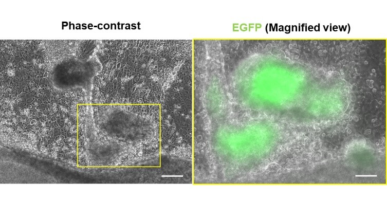
Fig.2 Confirmation of reporter system.
After PITX2 expression was induced, the part of the aggregated cells showed green fluorescent signals.
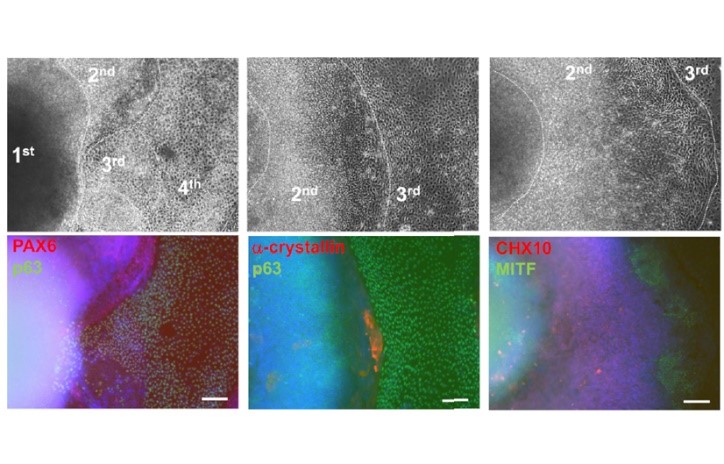
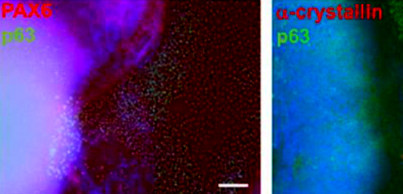
Fig.3 SEAM induction using reporter iPS cell line.
Normal SEAM structure was induced using the reporter iPS cell line: corneal epithelial cells (p63 /PAX6), lens cells (p63/a-crystallin), neuroretinal cells (CHX10), and retinal pigment epithelial cell (MITF).
The article, “Generation and validation of a PITX2-EGFP reporter line of human induced pluripotent stem cells enables isolation of periocular mesenchymal cells,” was published in Journal of Biological Chemistry at DOI: https://doi.org/10.1074/jbc.RA119.010713 .
Related links