
New microscopic technique clarifies early stage of osteogenesis
Living cells visualized at a nanoscale resolution
A research group at the Graduate School of Dentistry of Osaka University, in a joint research project with the National Institute of Advanced Industrial Science and Technology (AIST) and Lion Corporation, observed the process in which living osteoblasts form matrix vesicles (MVs) and secrete mineral precursors through MVs in the cell at a nanoscale resolution using a new microscopic technique.
They clarified that MVs formed in the cell were transported via lysosomes, which digest and dispose of unwanted substances in the cell, and were secreted by exocytosis. It was thought that mineralization depended on secretion of MVs with diameters of 30–300 nm, but the mechanism of how MVs were produced and secreted remained largely unknown.
With an electronic microscope capable of observing microscopic substances in the order of nanometers (one billionth of a meter), it was impossible to directly observe living cells because a specimen was placed in a vacuum sample chamber after sample fixation. It was also difficult to observe microscopic substances using an optical microscope that allowed one to directly observe living cells. Thus, detailed analysis of the mechanism did not advance and the description of the early stage of bone formation was based on hypotheses even in specialized books. Since the first report on the observation of MVs in 1967, the clarification of its formation and secretion process had not made progress.
In this study, in order to observe living cultured cells at high resolution to clarify the process of formation and secretion of MVs, the researchers used scanning electron-assisted dielectric microscopy (SE-ADM) developed by the AIST and a fluorescence optical microscope with super-high resolution. With SE-ADM, it’s possible to directly observe microscopic substances in living cells. They visualized MVs in living cells, clarifying that accumulated MVs in intact osteoblasts were transported via lysosomes and secreted by exocytosis.
The results will deepen the understanding of basic mechanisms of the formation of hard tissues, such as bones and teeth, leading to the clarification of conditions of diseases, such as osteoporosis and gum diseases, as well as the development of their treatment methods. SE-ADM that allows one to directly observe cultured cells as well as various microscopic substances in a solution will be used in various fields.
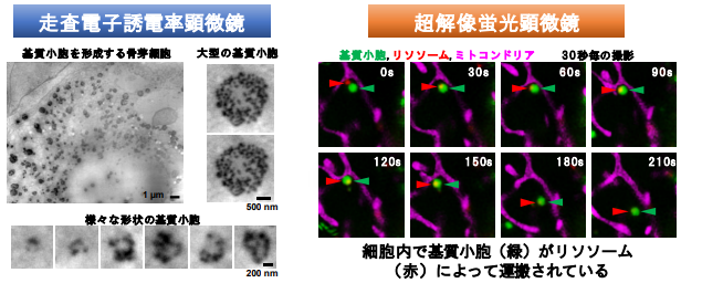
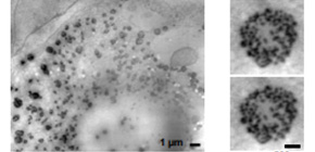
Figure 1
The article, “Osteoblastic lysosome plays a central role in mineralization,” was published in Science Advances at DOI: https://doi.org/10.1126/sciadv.aax0672 .
Related links