
Bright and Stable: New Acid-Tolerant Green Fluorescent Protein for Bioimaging
Osaka University researchers develop new green fluorescent protein that can withstand low pH environment for imaging of acidic organelles
Visualizing cellular components and processes at the molecular level is important for understanding the basis of any biological activity. Fluorescent proteins (FPs) are one of the most useful tools for investigating intracellular molecular dynamics.
However, FPs have usage limitations for imaging in low pH environments, such as in acidic organelles, including endosomes, lysosomes, and plant vacuoles. In environments of pH less than 6, most FPs lose their brightness and stability due to their neutral p K a. p K a is the measure of acid strength; the smaller the p K a is, the more acidic the substance is.
“Although there are reports of several acid-tolerant green FPs (GFPs), most have serious drawbacks. Furthermore, there is a lack of acid-tolerant GFPs that are practically applicable to bioimaging,” says Hajime Shinoda, lead author of an Osaka University study that aimed to design acid-tolerant monomeric GFP that is practically applicable to live-cell imaging in acidic organelles. “In the current study, we developed an acid-tolerant GFP. We called it Gamillus.”
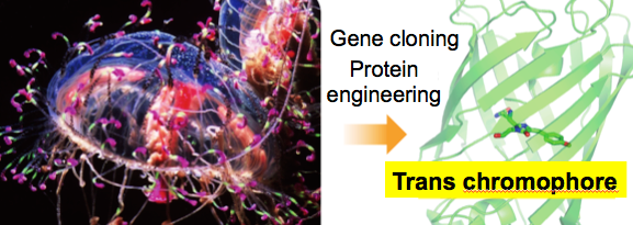
Gamillus is a GFP cloned from Olindias fo rmosa (flower hat jellyfish) and exhibits superior acid tolerance (p K a=3.4) and nearly twice as much brightness compared with the reported GFPs. The fluorescence spectrum is constant between pH4.5 and 9.0, which falls between the intracellular range in most cell types. X-ray crystallography (a technique used for determining the atomic and molecular structure of a crystal, in this case, a Gamillus crystal) and point mutagenesis suggest the acid tolerance of Gamillus is attributed to stabilization of deprotonation in its chemical structure. The findings were published in Cell Chemical Biology .
“The applicability of Gamillus as a molecular tag was shown by the correct localization pattern of Gamillus fusions in a variety of cellular structures, including ones that are difficult to target,” corresponding author Takeharu Nagai says. “We believe Gamillus can be a powerful molecular tool for investigating unknown biological phenomena involving acidic organelles, such as autophagy.”
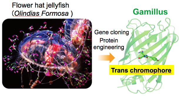
Figure 1. A schematic of development of Gamillus. Gene of the fluorescent protein was cloned from tentacles of flower hat jellyfish, and was engineered to improve the monomeric property and the brightness. (The flower hat jellyfish was provided by Mr. Kamoizumi in Kamo Aquarium, Yamagata, Japan.) (credit: Kamo Aquarium and Osaka University)
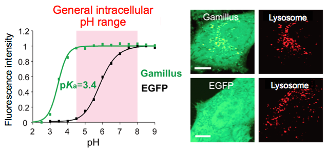
Figure 2. (Left) pH-dependent fluorescence property of Gamillus and EGFP. (Right) Fluorescence images of HeLa cells expressing Gamillus or EGFP. Gamillus enables fluorescence observation of protein migration to lysosomes caused by macroautophagy. Scale bar, 10 μm. (credit: Osaka University)
To learn more about this research, please view the full research report entitled "Acid-tolerant monomeric GFP from Olindias formosa " at this page of Cell Chemical Biology .
Related links