
Next-generation vascular endoscopic catheter with head-mounted image sensor developed through Osaka University-Panasonic Collaboration
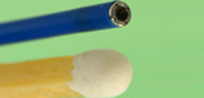
A group of researchers from Osaka University and Panasonic Corporation (Kadoma, Osaka) has launched the development of medical devices for observing blood vessels through industry-academy collaboration in the fields of medicine and engineering since 2013. They have succeeded in the commercialization of a vascular endoscopic catheter with a head-mounted image sensor. This vascular endoscopic catheter of 1.8mm diameter takes color photographs of about 480,000 pixels.
As medical devices for observing inside blood vessels, such as intravascular ultrasound (IVUS) and optical coherence tomography (OCT), provide blood vessel cross-section in monochrome images; however, medical staff in clinical practice wanted to provide treatment while viewing interior of blood vessels in real time.
The vascular endoscopic catheter developed by this group shows the blood vessel interiors in colored images using an image sensor at the distal end of the catheter.
To put this catheter into practical use, the group needed to achieve (1) high-precision processing technology to attach a small image sensor on the head of the thin tubular catheter to be inserted into the blood vessel and (2) technology to control the sensor and catheter to construct high-quality images.
A group of researchers led by former Osaka University Professor NANTO Shinsuke and Specially Appointed Assistant Professor OKAYAMA Keita at the Global Center for Medical Engineering and Informatics, Osaka University, came across technology of Panasonic Corp. at the MEDICA, World Forum for Medicine held in Düsseldorf in November 2013. Thinking that this technology could be used for their vascular endoscopic catheter under consideration, they laid out the framework for joint study shortly after returning to Japan.
The catheter they developed will provide medical staff not only with information about lesions in normal blood vessel treatment, but also with real-time information on the lesion to be treated in difficult-to-treat cases such as total occulusion lesions, which have recently been on the rise. The catheter has enabled detailed observation of arterial sclerosis and calcification in peripheral blood vessels, as well as blood clot formation and conditions after stent implantation. In addition to information on lesions necessary for their treatment, it will also provide useful information for evaluation of new drugs and stents.
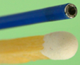
Figure 1
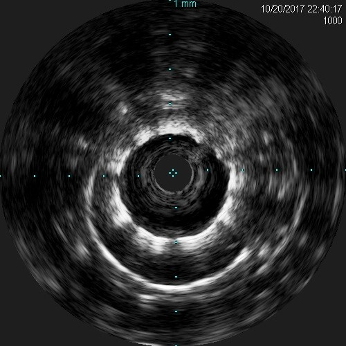
Figure 2
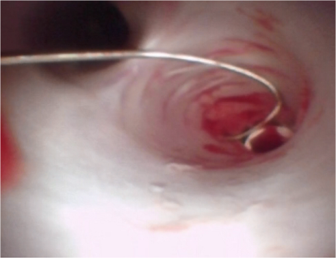
Figure 3
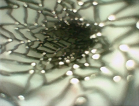
Figure 4
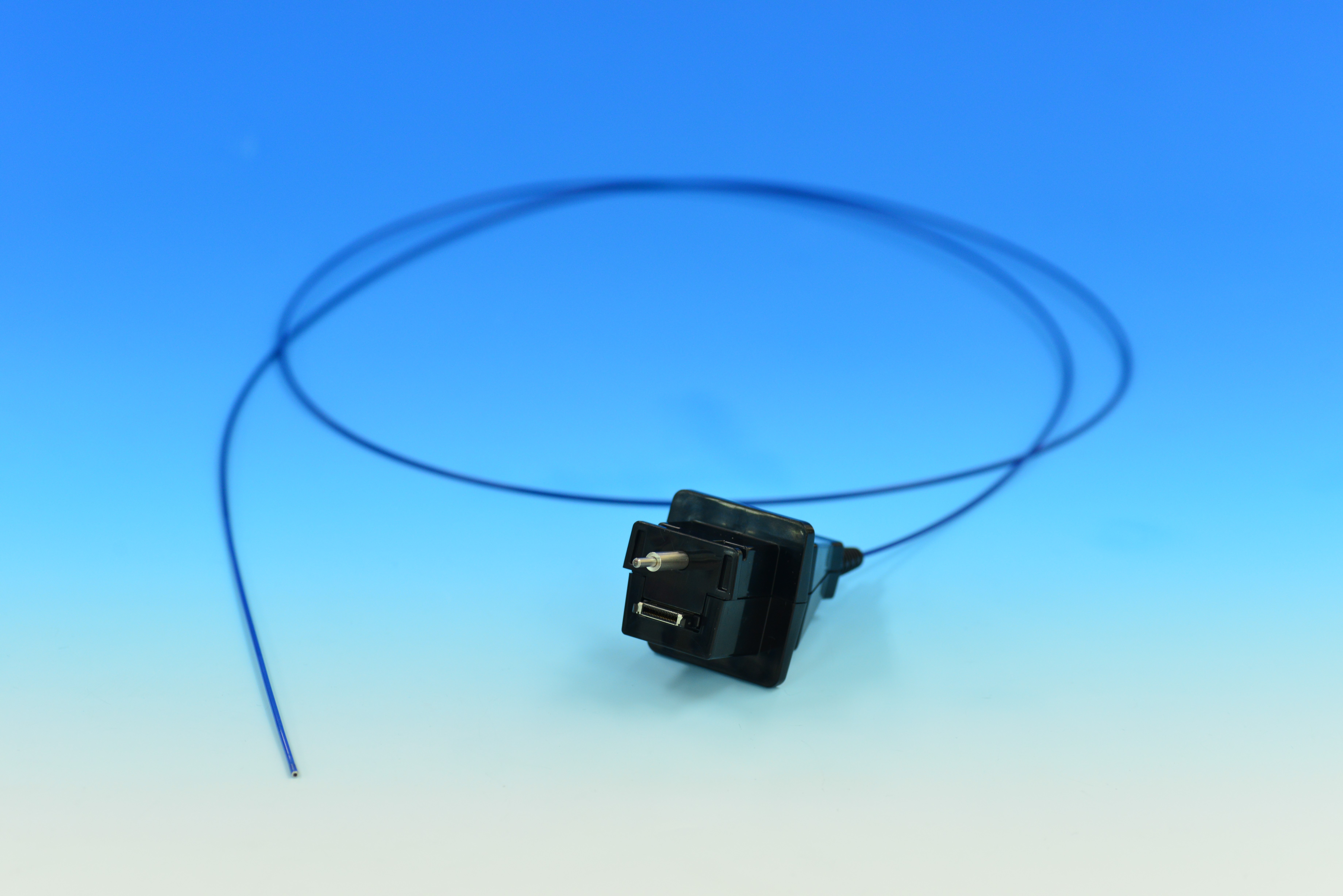
Figure 5
Related links
- Advanced Cardiovascular Therapeutics, Graduate School of Medicine, Osaka University (link in Japanese)
- Global Center for Medical Engineering and Informatics, Osaka University (link in Japanese)