
Mechanism for operating molecular motors facilitating transport of intracellular cargos clarified
Structures of the motion of “dynein”, a motor protein, were visualized!
A research group of Japanese universities, RIKEN and Leeds University in the UK succeeded in directly observing dynein, a motor protein facilitating transport of intracellular cargos, by using a low temperature electron microscope.
If the visualization of mutant dynein, which is related to various diseases, is achieved, it is anticipated that it will lead to the clarification of causes of neural diseases and growth disorder and the development of their treatments. It is also expected that it will lead to the elucidation of causes of cancer and applications to cancer treatment.
This research was featured in the electronic version of Nature Communications (UK) on Monday, September 14, 2015, Japan time.
Cells in our body have an efficient system for transporting various intracellular cargos such as nuclei, mitochondria, RNA, and proteins to the right place at the right time. This transport system is important for us to live. It is thought that if even a part of the function is lost, the body will develop cranial nerve diseases, developmental anomalies, and cancer.
The key players of a system to transport intracellular cargos are two protein groups called “molecular motors”: kinesin and dynein. Research on kinesin, a driving force behind the transport to the periphery of a cell (toward the microtubule plus end) has made progress to the point where the mechanism of movement can be discussed at an atomic level. On the contrary, the mechanism of dynein responsible for transport to the center of a cell (toward the microtubule minus end) had not been known in spite of energetic researchers’ efforts.
One of the major reasons for the prevention of the development of research on dynein was a lack of information about its structure. In recent years, the atomic structure of the core domain (motor domain) of dynein has been elucidated through X-ray crystallography* 2 . However, it was technologically difficult to catch the image of dynein moving along microtubules, which was most important in understanding dynein’s mechanism.
A group of researchers including IMAI Hiroshi, Assistant Professor, Faculty of Science and Engineering, Chuo University (previously worked at University of Leeds), KON Takahide, Professor, Graduate School of Science, Osaka University, SHIMA Tomohiro, Researcher, RIKEN (currently Assistant Professor, Graduate School of Science, The University of Tokyo), in cooperation with Dr. Stan A. Burgess and Professor Peter Knight University of Leeds (UK), succeeded in observing the structure of dyneins moving along microtubules for the first time through a low temperature electronic microscope. This group’s achievement was realized through the use of modified dyneins specially designed for this project (Figure 1), the optimization of conditions for observing through a low temperature electronic microscope over 6 years, and the application of a new image analysis method.
Important findings obtained in this project:
- 1. Two structures that dyneins take when they move along microtubules (Figure 2)
- 2. Dyneins move along microtubules using their two heads (motor area). Basic structural information necessary for understanding their motion mechanism in respective structures -- the directions to which the two heads face, their relative positional relationship, and the angle the two bond to the microtubule. (Figure 3)
- 3. Dyneins move along microtubules up to 20nm*4 while shaking in a position bound to the microtubule. (Figure 3)
Since the two major structures stated in item 1 were present in the same ratio, it is thought that dyneins move in these two structures alternatively. This way of moving is totally different from those of other molecule motors (kinesin and myosin) on which research has been conducted. It was found that dyneins facilitate transport of intracellular cargos in a unique way.
This group succeeded in directly observing the structure of molecular motors responsible for movement along microtubles in motion through a low temperature microscope for the first time in the world, which can be said to be a great technical innovation.

Figure 1. Diagram of the intact dynein being present in the cells, and the genetically modified dynein used in this study. Dynein is made of a single long polypeptide, consisting of three parts; (1) ~13-nm diameter ring, which hydrolyzes ATP, the chemical energy in the cells, (2) the extension structure (10 nm long with 2 nm diameter) from the ring, which is called the “stalk” (the tip of the stalk contains microtubule-binding site), and (3) the tail structure, which connects two rings. The (minimum) motor domain consists of the ring and the stalk without the tail.

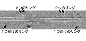
Figure 2. A cryo-electron micrograph taken at a freezing temperature of rapidly frozen dynein moving along microtubules. The long filament in the middle of the micrograph is the microtubule.
Several molecules of dynein can be seen as they bind to the microtubule. We observed mainly two structures: (1) the structure with two visible rings, and (2) the structure which looked like one visible ring but actually containing two overlapping rings. Microtubule polarity: plus-end towards the left of the micrograph and minus-end towards the right.

Figure 3.
The structures of dynein after applying image processing to the cryo-electron micrographs (the left four images in the vertical column) and atomic model (the right two panels).
Left: Two rings look like a single ring but actually overlap each other in this view. These three images above were obtained by image averaging after alignment and classification of many images of the dynein molecules. The large image at the bottom shows the averaging of such images. The stalks bound to microtubules, pointing towards the minus end of the microtubules, but the binding angles were variable.
The binding angles are shown in the top left corner panel in a yellow colour. Our results show that dynein has a hinge near the microtubule-binding domain, and moves with large flexibility due to the hinge.
Right: An atomic model of the dynein shown in the left panels. This panels show that dynein has two rings even though it looks like a ring in the left panel by looking from the different views.

Figure 4. The dynein walking mechanism along microtubules newly obtained by the results of this study. We could interpret that dynein alternately takes the two major structures because the ratio between the structure of two separate rings and the structure of overlapping rings was 1:1. The top and the bottom drawings correspond to the two major structures. The two drawings in the middle of the figure show intermediate states of the two major structures, which could be predicted by combination of our study and other studies. This is the first study which shows the overlapping motor structure on its track (shown at the top) among the motor proteins.
Related link