
Development of new microscopy based on high-speed SRS spectral microscopy and modified ICA
Under the leadership of Assistant Professor Yasuyuki OZEKI , Professor Kazuyoshi ITOH , Professor Kiichi FUKUI , former graduate student Wataru UMEMURA , Specially Appointed Assistant Professor Kazuhiko SUMIMURA (all Graduate School of Engineering, Osaka University); Japan Science and Technology Agency, Chief Hiroyuki HASHIMOTO ; Dr. Youichi OTSUKA , and Dr. Hideya SATO (all Canon Inc.), and Professor Norihiko NISHIZAWA , Nagoya University, a group of researchers have developed a microscope capable of real-time imaging of living tissues without staining, using a laser capable of fast switching of wavelengths. This device and independently-developed analytical algorithm offer imaging of three-dimensional structures and different constituents of living tissues as if they were stained.
Abstract
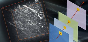
" To date, medical imaging of tissues has largely relied on time-consuming staining processes, and there is a need for rapid, label-free imaging techniques. Stimulated Raman scattering microscopy offers a three-dimensional, real-time imaging capability with chemical specificity. However, it can be difficult to differentiate between several constituents in tissues because their spectral characteristics can overlap. Furthermore, imaging speeds in previous multispectral stimulated Raman scattering imaging techniques were limited. Here, we demonstrate label-free imaging of tissues by 30 frames/s stimulated Raman scattering microscopy with frame-by-frame wavelength tunability. To produce multicolour images showing different constituents, spectral images were processed by modified independent component analysis, which can extract small differences in spectral features. We present various imaging modalities such as two-dimensional spectral imaging of rat liver, two-colour three-dimensional imaging of a vessel in rat liver, spectral imaging of several sections of intestinal villi in mouse, and in vivo spectral imaging of mouse ear skin. "
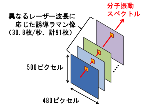
Figure 1

Figure 2
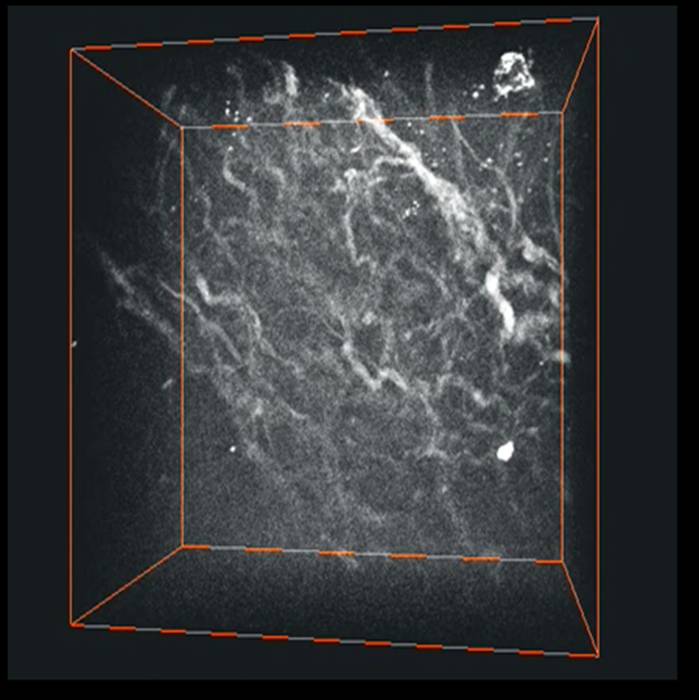
Figure 3

Figure 4
To learn more about this research, please read the full research report entitled " High-speed molecular spectral imaging of tissue with stimulated Raman scattering " on this page at the Nature Photonics website.
Related links