
Successful observation of cortical interneuron migration in living mouse embryos
contributes to understanding of brain formation in fetal development
Under the leadership of YANAGIDA Mitsutoshi , Researcher, and MURAKAMI Fujio , Professor at the Graduate School of Frontier Biosciences, Osaka University, a group of researchers have clarified cortical interneuron migration in living mouse embryos while keeping embryo umbilical cords attached to the mothers.
The neuron network of the brain is formed through the connection of neurons; however, this network is not formed in the brain development stage. In the development stage, neurons start migrating after the birth. After they reach locations they function as neurons, they then mature, forming neuron networks. Interneuron migration is important in the formation of brain functions; however, observation of such has been possible only in cultivated invitro neurons that had been taken from fetuses.
This group of researchers succeeded in recording the brain interneuron migration in living mouse embryos over 13 hours in stable conditions, clarifying how neurons moved ahead and changed directions.
Abstract
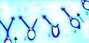
"Precisely arranged cytoarchitectures such as layers and nuclei depend on neuronal migration, of which many in vitro studies have revealed the mode and underlying mechanisms. However, how neuronal migration is achieved in vivo remains unknown. Here we established an imaging system that allows direct visualization of cortical interneuron migration in living mouse embryos. We found that during nucleokinesis, translocation of the Golgi apparatus either precedes or occurs in parallel to that of the nucleus, suggesting the existence of both a Golgi/centrosome-dependent and -independent mechanism of nucleokinesis. Changes in migratory direction occur when the nucleus enters one of the leading process branches, which is accompanied by the retraction of other branches. The nucleus occasionally swings between two branches before translocating into one of them, the occurrence of which is most often preceded by Golgi apparatus translocation into that branch. These in vivo observations provide important insight into the mechanisms of neuronal migration and demonstrate the usefulness of our system for studying dynamic events in living animals."
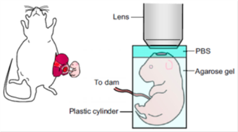
Figure 1
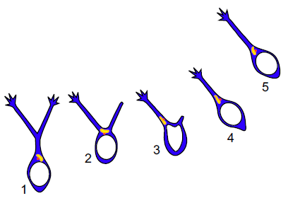
Figure 2
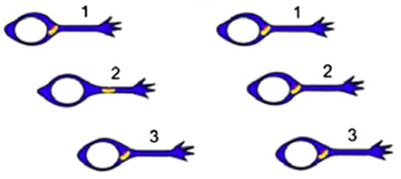
Figure 3
To learn more about this research, please read the full research report entitled "Dynamics of the leading process, nucleus, and Golgi apparatus of migrating cortical interneurons in living mouse embryos" on this page at the PNAS website.
Related link