
Correlative light-electron microscopy
an important step in the clarification of synaptic mechanisms
Under the leadership of Professor Jun-ichi TAKAGI , Dr. Hiroki TANAKA , and Associate professor Kenji IWASAKI at the Institute for Protein Research, Osaka University, and in cooperation with Yokohama City University, a group of researchers have succeeded in observing architecture of cell adhesion mediated by polymorphic synaptic adhesion molecules neurexin and neuroligin.
In this research, correlative light-electron microscopy combining techniques with different resolution capabilities -- crystallization and preliminary X-ray analysis (angstrom), electronic microscope (nanometer), optical microscope (micrometer) -- confirmed sheet formation at synapses that had been thought to be simple junctions between neurons.
This group achievement suggests that there are different types of synapses and it is expected that sheet formation at synapses may affect the regulation of neural networks. Obtaining a method for examining molecular mechanisms of synapse formation will be an important step toward the clarification of synaptic mechanisms.
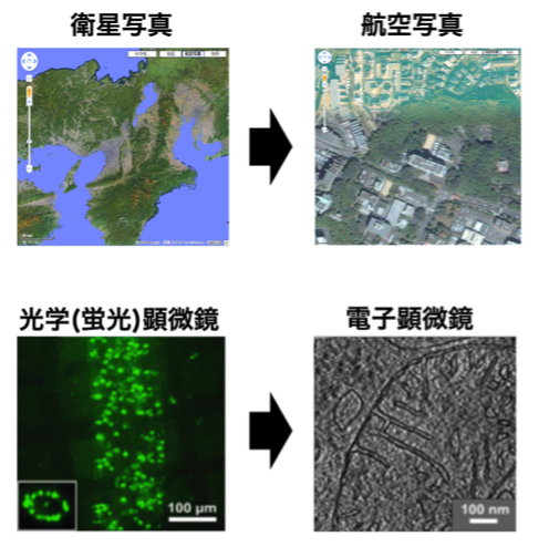
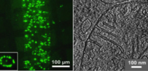
Figure 1
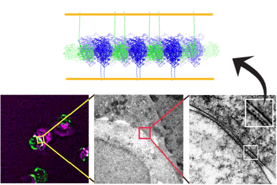
Figure 2
To learn more about this important discovery, please read the full research report entitled " Higher-Order Architecture of Cell Adhesion Mediated by Polymorphic Synaptic Adhesion Molecules Neurexin and Neuroligin " available at this page at the Cell Reports website.
Related link :