

Clarifying the Brain Mechanism for Stereoscopic Acuity
Synopsis
Under the leadership of Professor FUJITA Ichiro and Professor SHIOZAKI Hiroshi of the Graduate School of Frontier Biosciences , Osaka University, a team of researchers succeeded in identifying the area of the brain responsible for fine stereoscopic depth perception [stereoacuity] in humans and monkeys.
Further understanding of the mechanism providing information about depth perception in this area of the brain and greater understanding of the ways in which the brain processes visual information will be useful in the development of 3D technology that reduces eye strain and in the development of more natural stereoscopic images.
Background
In animals whose eyes are located on the same plane pointing straight ahead, the image from the right eye and the image from the left eye differ slightly. By measuring the level of this disparity, the brain is able to calculate the relative depth of object being viewed thereby providing the animal with stereoscopic vision.*1
In primates such as humans and monkeys, the level of stereoacuity is especially acute. For example, if two needles 2 meters from the viewer differ in distance by only 4 mm, humans and monkeys are able to tell which needle is closer or farther. This ability is referred to as fine stereoscopic acuity or stereoacuity, but which area of the brain is responsible for this ability has been unknown.
Findings
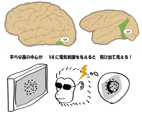
This group was able to clarify that the V4 area of the brain is responsible for fine stereoscopic acuity. *2
Visual areas make up more than half of the primate cerebral cortex and consist of more than 30 areas. This group analyzed the electric activities in the V4 area in monkeys wearing a pair of special stereo eyeglasses. As a result, the activity patterns in V4 cells were found to coincide with the depth perception of the monkeys.
Furthermore, this group demonstrated that the depth perception of the monkeys was affected when V4 cells activity was artificially induced. For example, by stimulating cell activity to convey information that an object has been placed in front, the monkeys mistook a flat surface for a protruding one. In this way, this group demonstrated that changes in cell activity in the V4 area caused the monkeys' depth perception to change. Formerly the relationship between the area of the parietal lobe and depth perception was mainly examined. This examination of the V4 area in the temporal lobe has clarified the area of the brain responsible for stereoscopic acuity.
Future development
This group's research has clarified how the brain is able to achieve fine stereoscopic depth perception and deepened understanding of the mechanism in the brain's stereoscopic vision. Further understanding of the nature of V4 cells will lead to design guidelines for 3D technology.*3
Such guidelines will help create more natural 3D images when using 3D CG software and greatly alleviate undue eye strain (pain at the back of the eye, headache, and/or nausea) caused by viewing unnatural stereoscopic images.
*1
The slight difference between the viewpoints of your two eyes is called binocular disparity. By means of binocular disparity and the distance between two eyes, the distance of objects can be determined by triangulation. The brain uses binocular disparity to obtain depth information and thus we are able to see objects in three dimensions -- binocular stereopsis. We can view the object in three dimensions with only one eye, but once we view it with both eyes, actual stereoscopic spatial observation is possible. This is a stereoscopic vision.
*2
Since the 1970s, it has been thought that stereoscopic vision consisted of 2 different abilities: coarse stereopsis and fine stereopsis. However, some researchers thought that coarse stereopsis and fine stereopsis were differing aspects of a single ability. These two views have competed for a long time in the field of perceptual psychology. In the 2000s, encephalous scientific evidence that the middle temporal visual area (MT) in the cerebral cortex of the macaque was related to coarse stereopsis but not to fine stereopsis was obtained. Now with this research clarifying that the V4 area is responsible for fine stereopsis, it has been confirmed that coarse stereopsis and fine stereopsis are two different abilities with two different areas of the brain responsible for them.
*3
It is not true that the greater the binocular disparity the greater the stereoscopic effect. The area of the brain responsible for fine stereopsis (as opposed to coarse stereopsis) senses planes with differing depths. Current 3D images are created with little regard for the fact that fine binocular disparity plays an important role in sensing the depth of a plane and unnecessarily large binocular disparity is given to images. Because of this, 3D scenography does not look natural, occasionally causing great eye strain. Further clarification of the brain mechanism of binocular streopsis will provide useful hints in the development of higher quality 3D image technology.